 Movie
Movie Controller

[English] 日本語
 Yorodumi
- PDB-2am9: Crystal structure of human androgen receptor ligand binding domai... -
+ Open data

- Basic information
| Entry | Database: PDB / ID: 2am9 | ||||||
|---|---|---|---|---|---|---|---|
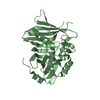
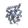
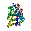
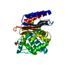
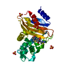
| Title | Crystal structure of human androgen receptor ligand binding domain in complex with testosterone | ||||||
 Components Components | Androgen receptor | ||||||
 Keywords Keywords | Hormone/Growth Factor Receptor / Nuclear Receptor / Androgen Receptor / Ligand Binding Domain / Testosterone / Agonist / Hormone-Growth Factor Receptor COMPLEX | ||||||
| Function / homology |  Function and homology information Function and homology informationmale somatic sex determination / activation of prostate induction by androgen receptor signaling pathway / lateral sprouting involved in mammary gland duct morphogenesis / POU domain binding / negative regulation of integrin biosynthetic process / regulation of developmental growth / male genitalia morphogenesis / positive regulation of integrin biosynthetic process / tertiary branching involved in mammary gland duct morphogenesis / animal organ formation ...male somatic sex determination / activation of prostate induction by androgen receptor signaling pathway / lateral sprouting involved in mammary gland duct morphogenesis / POU domain binding / negative regulation of integrin biosynthetic process / regulation of developmental growth / male genitalia morphogenesis / positive regulation of integrin biosynthetic process / tertiary branching involved in mammary gland duct morphogenesis / animal organ formation / androgen binding / Leydig cell differentiation / regulation of systemic arterial blood pressure / epithelial cell morphogenesis / prostate gland growth / epithelial cell differentiation involved in prostate gland development / non-membrane-bounded organelle assembly / positive regulation of epithelial cell proliferation involved in prostate gland development / prostate gland epithelium morphogenesis / intracellular receptor signaling pathway / cellular response to testosterone stimulus / RNA polymerase II general transcription initiation factor binding / positive regulation of insulin-like growth factor receptor signaling pathway / positive regulation of transcription by RNA polymerase III / positive regulation of intracellular estrogen receptor signaling pathway / cellular response to steroid hormone stimulus / morphogenesis of an epithelial fold / seminiferous tubule development / androgen receptor signaling pathway / RUNX2 regulates osteoblast differentiation / single fertilization / mammary gland alveolus development / intracellular estrogen receptor signaling pathway / cellular response to estrogen stimulus / regulation of protein localization to plasma membrane / estrogen response element binding / intracellular steroid hormone receptor signaling pathway / positive regulation of phosphorylation / HSP90 chaperone cycle for steroid hormone receptors (SHR) in the presence of ligand / steroid binding / insulin-like growth factor receptor signaling pathway / epithelial cell proliferation / molecular condensate scaffold activity / G protein-coupled receptor activity / positive regulation of cell differentiation / negative regulation of extrinsic apoptotic signaling pathway / SUMOylation of intracellular receptors / Activated PKN1 stimulates transcription of AR (androgen receptor) regulated genes KLK2 and KLK3 / multicellular organism growth / beta-catenin binding / transcription coactivator binding / positive regulation of miRNA transcription / Nuclear Receptor transcription pathway / nuclear receptor activity / negative regulation of epithelial cell proliferation / male gonad development / MAPK cascade / cell-cell signaling / positive regulation of NF-kappaB transcription factor activity / ATPase binding / spermatogenesis / DNA-binding transcription activator activity, RNA polymerase II-specific / in utero embryonic development / RNA polymerase II-specific DNA-binding transcription factor binding / transcription by RNA polymerase II / positive regulation of MAPK cascade / molecular adaptor activity / transcription cis-regulatory region binding / DNA-binding transcription factor activity, RNA polymerase II-specific / Ub-specific processing proteases / nuclear speck / RNA polymerase II cis-regulatory region sequence-specific DNA binding / DNA-binding transcription factor activity / negative regulation of cell population proliferation / signaling receptor binding / chromatin binding / chromatin / positive regulation of cell population proliferation / positive regulation of gene expression / positive regulation of DNA-templated transcription / enzyme binding / negative regulation of transcription by RNA polymerase II / signal transduction / positive regulation of transcription by RNA polymerase II / protein-containing complex / zinc ion binding / nucleoplasm / nucleus / plasma membrane / cytosol / cytoplasmSimilarity search - Function | ||||||
| Biological species |  Homo sapiens (human) Homo sapiens (human) | ||||||
| Method | X-RAY DIFFRACTION / MOLECULAR REPLACEMENT / Resolution: 1.64 Å | ||||||
 Authors Authors | Pereira de Jesus-Tran, K. / Cote, P.-L. / Cantin, L. / Blanchet, J. / Labrie, F. / Breton, R. | ||||||
 Citation Citation | Journal: Protein Sci. / Year: 2006 Title: Comparison of crystal structures of human androgen receptor ligand-binding domain complexed with various agonists reveals molecular determinants responsible for binding affinity. Authors: Pereira de Jesus-Tran, K. / Cote, P.-L. / Cantin, L. / Blanchet, J. / Labrie, F. / Breton, R. | ||||||
| History |
|
- Structure visualization
| Structure viewer | Molecule: MolmilJmol/JSmol |
|---|
- Downloads & links
-Download
| PDBx/mmCIF format | 2am9.cif.gz | 73.1 KB | Display | PDBx/mmCIF format |
|---|---|---|---|---|
| PDB format | pdb2am9.ent.gz | 53 KB | Display | PDB format |
| PDBx/mmJSON format | 2am9.json.gz | Tree view | PDBx/mmJSON format | |
| Others |  Other downloads Other downloads |
-Validation report
| Arichive directory | https://data.pdbj.org/pub/pdb/validation_reports/am/2am9ftp://data.pdbj.org/pub/pdb/validation_reports/am/2am9 | HTTPS FTP |
|---|
-Related structure data


-Links
 PDBj
PDBj


- Assembly
| Deposited unit | 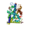
| ||||||||
|---|---|---|---|---|---|---|---|---|---|
| 1 |
| ||||||||
| Unit cell |
| ||||||||
| Details | The biological assembly is a monomer |
-Components
-Protein , 1 types, 1 molecules A
| #1: Protein | / Dihydrotestosterone receptor Mass: 30996.188 Da / Num. of mol.: 1 / Fragment: ligand binding domain Source method: isolated from a genetically manipulated source Source: (gene. exp.) Homo sapiens (human) / Gene: AR, DHTR, NR3C4 / Plasmid: pGEX / Production host:  Escherichia coli (E. coli) / Strain (production host): BL21(DE3)pLysS / References: UniProt: P10275 Escherichia coli (E. coli) / Strain (production host): BL21(DE3)pLysS / References: UniProt: P10275 |
|---|
-Non-polymers , 5 types, 227 molecules 


| #2: Chemical | ChemComp-SO4 / Sulfate Mass: 96.063 Da / Num. of mol.: 1 / Source method: obtained synthetically / Formula: SO4 Mass: 96.063 Da / Num. of mol.: 1 / Source method: obtained synthetically / Formula: SO4 |
|---|---|
| #3: Chemical | ChemComp-TES / Testosterone Mass: 288.424 Da / Num. of mol.: 1 / Source method: obtained synthetically / Formula: C19H28O2 / Comment: hormone*YM Mass: 288.424 Da / Num. of mol.: 1 / Source method: obtained synthetically / Formula: C19H28O2 / Comment: hormone*YM |
| #4: Chemical | ChemComp-DTT / Dithiothreitol Mass: 154.251 Da / Num. of mol.: 1 / Source method: obtained synthetically / Formula: C4H10O2S2 Mass: 154.251 Da / Num. of mol.: 1 / Source method: obtained synthetically / Formula: C4H10O2S2 |
| #5: Chemical | ChemComp-GOL / Glycerol Mass: 92.094 Da / Num. of mol.: 1 / Source method: obtained synthetically / Formula: C3H8O3 Mass: 92.094 Da / Num. of mol.: 1 / Source method: obtained synthetically / Formula: C3H8O3 |
| #6: Water | ChemComp-HOH / WaterMass: 18.015 Da / Num. of mol.: 223 / Source method: isolated from a natural source / Formula: H2O |
-Experimental details
-Experiment
| Experiment | Method: X-RAY DIFFRACTION / Number of used crystals: 1 |
|---|
- Sample preparation
| Crystal | Density Matthews: 1.87 Å3/Da / Density % sol: 33.8 % |
|---|---|
| Crystal grow | Temperature: 293 K / Method: vapor diffusion, hanging drop / pH: 7.6 Details: Bicine, Na/K tartrate, MgSO4, pH 7.6, VAPOR DIFFUSION, HANGING DROP, temperature 293K |
-Data collection
| Diffraction | Mean temperature: 100 K |
|---|---|
| Diffraction source | Source: ROTATING ANODE / Type: RIGAKU RU200 / Wavelength: 1.5418 Å |
| Detector | Type: RIGAKU RAXIS II / Detector: IMAGE PLATE / Date: Jan 10, 2005 / Details: mirrors |
| Radiation | Protocol: SINGLE WAVELENGTH / Monochromatic (M) / Laue (L): M / Scattering type: x-ray |
| Radiation wavelength | Wavelength: 1.5418 Å / Relative weight: 1 |
| Reflection | Resolution: 1.64→19.23 Å / Num. obs: 30947 / % possible obs: 96.8 % / Observed criterion σ(F): 1 / Observed criterion σ(I): 1 / Redundancy: 10.5 % / Biso Wilson estimate: 29.42 Å2 / Rmerge(I) obs: 0.062 / Rsym value: 0.06 / Net I/σ(I): 22.02 |
| Reflection shell | Resolution: 1.64→1.7 Å / % possible obs: 77 % / Redundancy: 3.5 % / Rmerge(I) obs: 0.504 / Mean I/σ(I) obs: 2.98 / Num. measured obs: 8529 / Num. unique all: 2434 / Num. unique obs: 2434 / Rsym value: 0.433 / % possible all: 77 |
- Processing
| Software |
| ||||||||||||||||||||||||||||
|---|---|---|---|---|---|---|---|---|---|---|---|---|---|---|---|---|---|---|---|---|---|---|---|---|---|---|---|---|---|
| Refinement | Method to determine structure: MOLECULAR REPLACEMENT Starting model: hARLBD-DHT (in deposition) Resolution: 1.64→19.23 Å / Isotropic thermal model: Isotropic / Cross valid method: THROUGHOUT / σ(F): 0 / Stereochemistry target values: Engh & Huber
| ||||||||||||||||||||||||||||
| Solvent computation | Bsol: 62.67 Å2 | ||||||||||||||||||||||||||||
| Displacement parameters | Biso mean: 27.382 Å2
| ||||||||||||||||||||||||||||
| Refine analyze | Luzzati coordinate error obs: 0.24 Å / Luzzati d res low obs: 5 Å / Luzzati sigma a obs: 0.32 Å | ||||||||||||||||||||||||||||
| Refinement step | Cycle: LAST / Resolution: 1.64→19.23 Å
| ||||||||||||||||||||||||||||
| Refine LS restraints |
| ||||||||||||||||||||||||||||
| LS refinement shell | Resolution: 1.64→1.74 Å / Rfactor Rfree error: 0.026
| ||||||||||||||||||||||||||||
| Xplor file |
|
