Movie
Movie Controller

[English] 日本語
 Yorodumi
- PDB-2agn: Fitting of hepatitis C virus internal ribosome entry site domains... -
+ Open data

- Basic information
| Entry | Database: PDB / ID: 2agn | ||||||
|---|---|---|---|---|---|---|---|
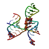
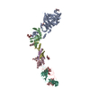
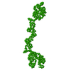
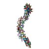
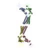
| Title | Fitting of hepatitis C virus internal ribosome entry site domains into the 15 A Cryo-EM map of a HCV IRES-80S ribosome (H. sapiens) complex | ||||||
 Components Components |
| ||||||
 Keywords Keywords |  RNA / HCV / IRES RNA / HCV / IRES | ||||||
| Function / homology | RNA / RNA (> 10) Function and homology information Function and homology information | ||||||
| Biological species | synthetic construct (others) | ||||||
| Method | ELECTRON MICROSCOPY / single particle reconstruction / cryo EM / Resolution: 15 Å | ||||||
 Authors Authors | Boehringer, D. / Thermann, R. / Ostareck-Lederer, A. / Lewis, J.D. / Stark, H. | ||||||
 Citation Citation | Journal: Structure / Year: 2005 Title: Structure of the hepatitis C virus IRES bound to the human 80S ribosome: remodeling of the HCV IRES. Authors: Daniel Boehringer / Rolf Thermann / Antje Ostareck-Lederer / Joe D Lewis / Holger Stark /  Abstract: Initiation of translation of the hepatitis C virus (HCV) polyprotein is driven by an internal ribosome entry site (IRES) RNA that bypasses much of the eukaryotic translation initiation machinery. ...Initiation of translation of the hepatitis C virus (HCV) polyprotein is driven by an internal ribosome entry site (IRES) RNA that bypasses much of the eukaryotic translation initiation machinery. Here, single-particle electron cryomicroscopy has been used to study the mechanism of HCV IRES-mediated initiation. A HeLa in vitro translation system was used to assemble human IRES-80S ribosome complexes under near physiological conditions; these were stalled before elongation. Domain 2 of the HCV IRES is bound to the tRNA exit site, touching the L1 stalk of the 60S subunit, suggesting a mechanism for the removal of the HCV IRES in the progression to elongation. Domain 3 of the HCV IRES positions the initiation codon in the ribosomal mRNA binding cleft by binding helix 28 at the head of the 40S subunit. The comparison with the previously published binary 40S-HCV IRES complex reveals structural rearrangements in the two pseudoknot structures of the HCV IRES in translation initiation. | ||||||
| History |
|
- Structure visualization
| Movie |
Movie viewer |
|---|---|
| Structure viewer | Molecule: MolmilJmol/JSmol |
- Downloads & links
-Download
| PDBx/mmCIF format | 2agn.cif.gz | 13.6 KB | Display | PDBx/mmCIF format |
|---|---|---|---|---|
| PDB format | pdb2agn.ent.gz | 6.9 KB | Display | PDB format |
| PDBx/mmJSON format | 2agn.json.gz | Tree view | PDBx/mmJSON format | |
| Others |  Other downloads Other downloads |
-Validation report
| Arichive directory | https://data.pdbj.org/pub/pdb/validation_reports/ag/2agnftp://data.pdbj.org/pub/pdb/validation_reports/ag/2agn | HTTPS FTP |
|---|
-Related structure data
| Related structure data | 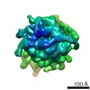 1138MC M: map data used to model this data C: citing same article ( |
|---|---|
| Similar structure data |
-Links
 PDBj
PDBj


- Assembly
| Deposited unit | 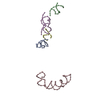
|
|---|---|
| 1 |
|
-Components
| #1: RNA chain | Mass: 9450.667 Da / Num. of mol.: 1 / Source method: obtained synthetically / Source: (synth.) synthetic construct (others) |
|---|---|
| #2: RNA chain | Mass: 9595.754 Da / Num. of mol.: 1 / Source method: obtained synthetically / Source: (synth.) synthetic construct (others) |
| #3: RNA chain | Mass: 24776.691 Da / Num. of mol.: 1 / Source method: obtained synthetically / Source: (synth.) synthetic construct (others) |
| #4: RNA chain | Mass: 4547.780 Da / Num. of mol.: 1 / Source method: obtained synthetically / Source: (synth.) synthetic construct (others) |
| #5: RNA chain | Mass: 17161.283 Da / Num. of mol.: 1 / Source method: obtained synthetically / Source: (synth.) synthetic construct (others) |
-Experimental details
-Experiment
| Experiment | Method: ELECTRON MICROSCOPY |
|---|---|
| EM experiment | Aggregation state: PARTICLE / 3D reconstruction method: single particle reconstruction |
- Sample preparation
| Component | Name: HCV IRES-80S ribosome complex / Type: RIBOSOME |
|---|---|
| Buffer solution | pH: 8.1 |
| Specimen | Embedding applied: NO / Shadowing applied: NO / Staining applied: NO / Vitrification applied: YES |
| Specimen support | Details: perforated corbon foil on a copper grid |
| Vitrification | Cryogen name: ETHANE / Details: plunging into liquid ethane |
- Electron microscopy imaging
| Microscopy | Model: FEI/PHILIPS CM200FEG / Date: Oct 1, 2003 |
|---|---|
| Electron gun | Electron source: FIELD EMISSION GUN / Accelerating voltage: 200 kV / Illumination mode: FLOOD BEAM |
| Electron lens | Mode: BRIGHT FIELDBright-field microscopy / Nominal magnification: 50000 X / Calibrated magnification: 50000 X / Nominal defocus max: 4000 nm / Nominal defocus min: 2000 nm / Cs: 2 mm |
| Specimen holder | Temperature: 77 K / Tilt angle max: 0 ° / Tilt angle min: 0 ° |
| Image recording | Electron dose: 20 e/Å2 / Film or detector model: GENERIC FILM / Details: Kodak SO-163 |
| Radiation | Protocol: SINGLE WAVELENGTH / Monochromatic (M) / Laue (L): M / Scattering type: x-ray |
| Radiation wavelength | Relative weight: 1 |
- Processing
| EM software |
| |||||||||||||||||||||||||||||||||||
|---|---|---|---|---|---|---|---|---|---|---|---|---|---|---|---|---|---|---|---|---|---|---|---|---|---|---|---|---|---|---|---|---|---|---|---|---|
| CTF correction | Details: phase reversal | |||||||||||||||||||||||||||||||||||
| Symmetry | Point symmetry: C1 (asymmetric) | |||||||||||||||||||||||||||||||||||
| 3D reconstruction | Method: angular reconstitution / Resolution: 15 Å / Num. of particles: 24100 / Nominal pixel size: 3.6 Å / Actual pixel size: 3.6 Å Details: This entry contains only phosphorus atom in the coordinate. Symmetry type: POINT | |||||||||||||||||||||||||||||||||||
| Atomic model building | Space: REAL | |||||||||||||||||||||||||||||||||||
| Atomic model building |
| |||||||||||||||||||||||||||||||||||
| Refinement step | Cycle: LAST
|