 Movie
Movie Controller

[English] 日本語
 Yorodumi
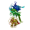
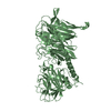
- PDB-2a75: Trypanosoma rangeli Sialidase In Complex With 2,3- Difluorosialic... -
+ Open data

- Basic information
| Entry | Database: PDB / ID: 2a75 | ||||||
|---|---|---|---|---|---|---|---|
| Title | Trypanosoma rangeli Sialidase In Complex With 2,3- Difluorosialic Acid (Covalent Intermediate) | ||||||
 Components Components | sialidase Neuraminidase Neuraminidase | ||||||
 Keywords Keywords | HYDROLASE / beta-propeller / covalent enzyme-intermediate complex / beta-sandwich | ||||||
| Function / homology |  Function and homology information Function and homology informationexo-alpha-(2->3)-sialidase activity / exo-alpha-(2->6)-sialidase activity / exo-alpha-(2->8)-sialidase activity / exo-alpha-sialidase / metabolic processSimilarity search - Function | ||||||
| Biological species |  Trypanosoma rangeli (eukaryote) Trypanosoma rangeli (eukaryote) | ||||||
| Method | X-RAY DIFFRACTION / SYNCHROTRON / FOURIER SYNTHESIS / Resolution: 1.95 Å | ||||||
 Authors Authors | Amaya, M.F. / Alzari, P.M. / Buschiazzo, A. | ||||||
 Citation Citation | Journal: J.Biol.Chem. / Year: 2006 Title: Structural and Kinetic Analysis of Two Covalent Sialosyl-Enzyme Intermediates on Trypanosoma rangeli Sialidase. Authors: Watts, A.G. / Oppezzo, P. / Withers, S.G. / Alzari, P.M. / Buschiazzo, A. | ||||||
| History |
| ||||||
| Remark 600 | HETEROGEN The covalent Sialo-Tyrosine adduct is the product of the reaction of 2,3-difluorosialic ...HETEROGEN The covalent Sialo-Tyrosine adduct is the product of the reaction of 2,3-difluorosialic acid with the enzyme | ||||||
| Remark 999 | SEQUENCE Authors indicate that the sequence in the database is incorrect |
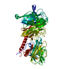
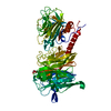
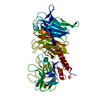
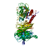
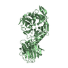
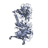
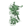
- Structure visualization
| Structure viewer | Molecule: MolmilJmol/JSmol |
|---|
- Downloads & links
-Download
| PDBx/mmCIF format | 2a75.cif.gz | 153.2 KB | Display | PDBx/mmCIF format |
|---|---|---|---|---|
| PDB format | pdb2a75.ent.gz | 115.8 KB | Display | PDB format |
| PDBx/mmJSON format | 2a75.json.gz | Tree view | PDBx/mmJSON format | |
| Others |  Other downloads Other downloads |
-Validation report
| Arichive directory | https://data.pdbj.org/pub/pdb/validation_reports/a7/2a75ftp://data.pdbj.org/pub/pdb/validation_reports/a7/2a75 | HTTPS FTP |
|---|
-Related structure data
| Related structure data |  2agsC 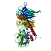 2fhrC 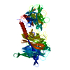 1n1sS S: Starting model for refinement C: citing same article ( |
|---|---|
| Similar structure data |


-Links
 PDBj
PDBj

- Assembly
| Deposited unit | 
| ||||||||
|---|---|---|---|---|---|---|---|---|---|
| 1 |
| ||||||||
| Unit cell |
| ||||||||
| Details | The biological unit is a monomer |
-Components
| #1: Protein | Neuraminidase Mass: 71205.203 Da / Num. of mol.: 1 Source method: isolated from a genetically manipulated source Source: (gene. exp.) Trypanosoma rangeli (eukaryote) / Plasmid: pTrcHisA / Production host:  Escherichia coli (E. coli) / Strain (production host): TOP10 / References: UniProt: O44049, exo-alpha-sialidase Escherichia coli (E. coli) / Strain (production host): TOP10 / References: UniProt: O44049, exo-alpha-sialidase | ||||
|---|---|---|---|---|---|
| #2: Chemical | Sulfate  Mass: 96.063 Da / Num. of mol.: 2 / Source method: obtained synthetically / Formula: SO4 Mass: 96.063 Da / Num. of mol.: 2 / Source method: obtained synthetically / Formula: SO4#3: Sugar | ChemComp-FSI / | 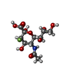  Type: D-saccharide, beta linking / Mass: 327.260 Da / Num. of mol.: 1 Type: D-saccharide, beta linking / Mass: 327.260 Da / Num. of mol.: 1Source method: isolated from a genetically manipulated source Formula: C11H18FNO9 #4: Water | ChemComp-HOH / | Water Mass: 18.015 Da / Num. of mol.: 615 / Source method: isolated from a natural source / Formula: H2O Mass: 18.015 Da / Num. of mol.: 615 / Source method: isolated from a natural source / Formula: H2O |
-Experimental details
-Experiment
| Experiment | Method: X-RAY DIFFRACTION / Number of used crystals: 1 |
|---|
- Sample preparation
| Crystal | Density Matthews: 2.7 Å3/Da / Density % sol: 54.4 % |
|---|---|
| Crystal grow | Temperature: 291 K / Method: vapor diffusion, hanging drop / pH: 6.5 Details: PEG 8000, ammonium sulfate, sodium cacodylate, pH 6.5, VAPOR DIFFUSION, HANGING DROP, temperature 291K |
-Data collection
| Diffraction | Mean temperature: 100 K |
|---|---|
| Diffraction source | Source: SYNCHROTRON / Site: ESRF  / Beamline: ID29 / Wavelength: 0.979158 Å / Beamline: ID29 / Wavelength: 0.979158 Å |
| Detector | Type: ADSC QUANTUM 210 / Detector: CCD / Date: Nov 2, 2003 |
| Radiation | Monochromator: Si 311 channel-cut / Protocol: SINGLE WAVELENGTH / Monochromatic (M) / Laue (L): M / Scattering type: x-ray |
| Radiation wavelength | Wavelength: 0.979158 Å / Relative weight: 1 |
| Reflection | Resolution: 1.9→46.63 Å / Num. all: 47608 / Num. obs: 47608 / % possible obs: 91.2 % / Observed criterion σ(F): 1 / Observed criterion σ(I): 1 / Redundancy: 2.9 % / Rmerge(I) obs: 0.111 / Rsym value: 0.111 / Net I/σ(I): 3.9 |
| Reflection shell | Resolution: 1.9→2 Å / % possible obs: 80.4 % / Redundancy: 2.6 % / Rmerge(I) obs: 0.3 / Mean I/σ(I) obs: 1.7 / Num. measured obs: 6972 / Num. unique all: 6972 / Rsym value: 0.3 / % possible all: 80.4 |
- Processing
| Software |
| ||||||||||||||||||||||||||||||||||||||||||||||||||||||||||||||||||||||||||||||||||||||||||
|---|---|---|---|---|---|---|---|---|---|---|---|---|---|---|---|---|---|---|---|---|---|---|---|---|---|---|---|---|---|---|---|---|---|---|---|---|---|---|---|---|---|---|---|---|---|---|---|---|---|---|---|---|---|---|---|---|---|---|---|---|---|---|---|---|---|---|---|---|---|---|---|---|---|---|---|---|---|---|---|---|---|---|---|---|---|---|---|---|---|---|---|
| Refinement | Method to determine structure: FOURIER SYNTHESIS Starting model: pdb entry 1N1S Resolution: 1.95→29.04 Å / Rfactor Rfree error: 0.004 / Occupancy max: 1 / Occupancy min: 0.5 / Isotropic thermal model: Isotropic / Cross valid method: THROUGHOUT / σ(F): 0 / Stereochemistry target values: Engh & Huber
| ||||||||||||||||||||||||||||||||||||||||||||||||||||||||||||||||||||||||||||||||||||||||||
| Solvent computation | Solvent model: CNS bulk solvent model / Bsol: 46.1351 Å2 / ksol: 0.364945 e/Å3 | ||||||||||||||||||||||||||||||||||||||||||||||||||||||||||||||||||||||||||||||||||||||||||
| Displacement parameters | Biso max: 56.5 Å2 / Biso mean: 11.58 Å2 / Biso min: 1.36 Å2
| ||||||||||||||||||||||||||||||||||||||||||||||||||||||||||||||||||||||||||||||||||||||||||
| Refine analyze |
| ||||||||||||||||||||||||||||||||||||||||||||||||||||||||||||||||||||||||||||||||||||||||||
| Refinement step | Cycle: LAST / Resolution: 1.95→29.04 Å
| ||||||||||||||||||||||||||||||||||||||||||||||||||||||||||||||||||||||||||||||||||||||||||
| Refine LS restraints |
| ||||||||||||||||||||||||||||||||||||||||||||||||||||||||||||||||||||||||||||||||||||||||||
| LS refinement shell | Refine-ID: X-RAY DIFFRACTION
|
