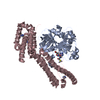
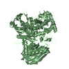
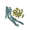
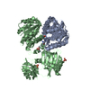
Mass: 29772.422 Da / Num. of mol.: 2 / Fragment: Thrombin heavy chain / Source method: isolated from a natural source / Source: (natural) Bos taurus (cattle) / References: UniProt: P00735, thrombin
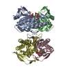
#3: Protein
Staphylocoagulase
Mass: 38541.992 Da / Num. of mol.: 2 / Fragment: active fragment 1-329 Source method: isolated from a genetically manipulated source Source: (gene. exp.) Staphylococcus aureus (bacteria) / Production host: Escherichia coli (E. coli) / References: UniProt: P17855
Mass: 22.990 Da / Num. of mol.: 2 / Source method: obtained synthetically / Formula: Na
-
Details
Nonpolymer details
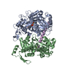
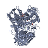
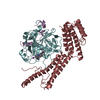
THE INHIBITOR IS COVALENTLY CONNECTED TO ACTIVE_SITE RESIDUES: 1) VIA A HEMIKETAL GROUP TO OG SER ...THE INHIBITOR IS COVALENTLY CONNECTED TO ACTIVE_SITE RESIDUES: 1) VIA A HEMIKETAL GROUP TO OG SER 195 IN CHAINS B AND F, 2) VIA A METHYLENE GROUP TO NE2 HIS 57 IN CHAINS B AND F.
Sequence details
THIS DIVERGENCE OF GLY FROM THE REPORTED SEQUENCE, PERHAPS ADDING THAT THIS VARIATION IS IRRELEVANT ...THIS DIVERGENCE OF GLY FROM THE REPORTED SEQUENCE, PERHAPS ADDING THAT THIS VARIATION IS IRRELEVANT FOR (PRO)THROMBIN BINDING, WHICH IS AN IMPORTANT POINT OF THIS ENTRY.
-
Experimental details
-
Experiment
Experiment
Method: X-RAY DIFFRACTION / Number of used crystals: 1
-
Sample preparation
Crystal
Density Matthews: 3.4 Å3/Da / Density % sol: 62.4 %
Crystal grow
Temperature: 298 K / Method: vapor diffusion, sitting drop / pH: 6.5 Details: 0.1M IMIDAZOLE, 0.2M AMMONIUM FORMATE, 12% POLYETHYLENE GLYCOL 4000, pH 6.5, VAPOR DIFFUSION, SITTING DROP, temperature 298K
In the structure databanks used in Yorodumi, some data are registered as the other names, "COVID-19 virus" and "2019-nCoV". Here are the details of the virus and the list of structure data.
Jan 31, 2019. EMDB accession codes are about to change! (news from PDBe EMDB page)
EMDB accession codes are about to change! (news from PDBe EMDB page)
The allocation of 4 digits for EMDB accession codes will soon come to an end. Whilst these codes will remain in use, new EMDB accession codes will include an additional digit and will expand incrementally as the available range of codes is exhausted. The current 4-digit format prefixed with “EMD-” (i.e. EMD-XXXX) will advance to a 5-digit format (i.e. EMD-XXXXX), and so on. It is currently estimated that the 4-digit codes will be depleted around Spring 2019, at which point the 5-digit format will come into force.
The EM Navigator/Yorodumi systems omit the EMD- prefix.
Related info.:Q: What is EMD? / ID/Accession-code notation in Yorodumi/EM Navigator
Yorodumi is a browser for structure data from EMDB, PDB, SASBDB, etc.
This page is also the successor to EM Navigator detail page, and also detail information page/front-end page for Omokage search.
The word "yorodu" (or yorozu) is an old Japanese word meaning "ten thousand". "mi" (miru) is to see.
Related info.:EMDB / PDB / SASBDB / Comparison of 3 databanks / Yorodumi Search / Aug 31, 2016. New EM Navigator & Yorodumi / Yorodumi Papers / Jmol/JSmol / Function and homology information / Changes in new EM Navigator and Yorodumi
 Movie
Movie Controller


 Open data
 Basic information
 Components
 Keywords
 Function and homology information
 fibrinogen binding /


 Authors
 Citation
 Structure visualization
 Downloads & links
 Other downloads
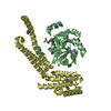


 PDBj
PDBj


 Assembly


 Type: D-saccharide, beta linking / Mass: 221.208 Da / Num. of mol.: 1

 Type: peptide-like
 Mass: 22.990 Da / Num. of mol.: 2 / Source method: obtained synthetically / Formula: Na
 Sample preparation
 Processing