 Movie
Movie Controller


+ Open data

- Basic information
| Entry | Database: PDB / ID: 1zxk | ||||||
|---|---|---|---|---|---|---|---|
| Title | Crystal Structure of Cadherin8 EC1 domain | ||||||
 Components Components | Cadherin-8 | ||||||
 Keywords Keywords | CELL ADHESION / cadherin / strand dimer | ||||||
| Function / homology |  Function and homology information Function and homology informationAdherens junctions interactions / cell-cell adhesion mediated by cadherin / calcium-dependent cell-cell adhesion via plasma membrane cell adhesion molecules / catenin complex / cell-cell junction assembly / adherens junction organization / regulation of synapse organization / homophilic cell adhesion via plasma membrane adhesion molecules / axon terminus / synaptic cleft ...Adherens junctions interactions / cell-cell adhesion mediated by cadherin / calcium-dependent cell-cell adhesion via plasma membrane cell adhesion molecules / catenin complex / cell-cell junction assembly / adherens junction organization / regulation of synapse organization / homophilic cell adhesion via plasma membrane adhesion molecules / axon terminus / synaptic cleft / response to cold / synaptic membrane / synaptic transmission, glutamatergic / adherens junction / cell morphogenesis / chemical synaptic transmission / cadherin binding / glutamatergic synapse / calcium ion binding / identical protein binding / plasma membraneSimilarity search - Function | ||||||
| Biological species |  Mus musculus (house mouse) Mus musculus (house mouse) | ||||||
| Method | X-RAY DIFFRACTION / SYNCHROTRON / MOLECULAR REPLACEMENT / Resolution: 2 Å | ||||||
 Authors Authors | Patel, S.D. / Ciatto, C. / Chen, C.P. / Bahna, F. / Arkus, N. / Schieren, I. / Rajebhosale, M. / Jessell, T.M. / Honig, B. / Price, S.R. / Shapiro, L. | ||||||
 Citation Citation | Journal: Cell(Cambridge,Mass.) / Year: 2006 Title: Type II cadherin ectodomain structures: implications for classical cadherin specificity. Authors: Patel, S.D. / Ciatto, C. / Chen, C.P. / Bahna, F. / Rajebhosale, M. / Arkus, N. / Schieren, I. / Jessell, T.M. / Honig, B. / Price, S.R. / Shapiro, L. | ||||||
| History |
|
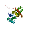
- Structure visualization
| Structure viewer | Molecule: MolmilJmol/JSmol |
|---|
- Downloads & links
-Download
| PDBx/mmCIF format | 1zxk.cif.gz | 53.3 KB | Display | PDBx/mmCIF format |
|---|---|---|---|---|
| PDB format | pdb1zxk.ent.gz | 38.2 KB | Display | PDB format |
| PDBx/mmJSON format | 1zxk.json.gz | Tree view | PDBx/mmJSON format | |
| Others |  Other downloads Other downloads |
-Validation report
| Arichive directory | https://data.pdbj.org/pub/pdb/validation_reports/zx/1zxkftp://data.pdbj.org/pub/pdb/validation_reports/zx/1zxk | HTTPS FTP |
|---|
-Related structure data
| Related structure data |  1zvnSC 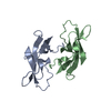 2a4cC 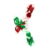 2a4eC  2a62C S: Starting model for refinement C: citing same article ( |
|---|---|
| Similar structure data |


-Links
 PDBj
PDBj
- Assembly
| Deposited unit | 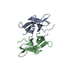
| ||||||||
|---|---|---|---|---|---|---|---|---|---|
| 1 |
| ||||||||
| Unit cell |
| ||||||||
| Components on special symmetry positions |
|
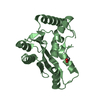
-Components
| #1: Protein | Mass: 11085.474 Da / Num. of mol.: 2 / Fragment: EC1 domain / Mutation: G1S Source method: isolated from a genetically manipulated source Source: (gene. exp.) Mus musculus (house mouse) / Gene: Cdh8 / Plasmid: pSMT3 / Production host:  Escherichia coli (E. coli) / References: UniProt: P97291 Escherichia coli (E. coli) / References: UniProt: P97291#2: Water | ChemComp-HOH / | Water Mass: 18.015 Da / Num. of mol.: 160 / Source method: isolated from a natural source / Formula: H2O Mass: 18.015 Da / Num. of mol.: 160 / Source method: isolated from a natural source / Formula: H2O |
|---|
-Experimental details
-Experiment
| Experiment | Method: X-RAY DIFFRACTION / Number of used crystals: 1 |
|---|
- Sample preparation
| Crystal | Density Matthews: 2.69 Å3/Da / Density % sol: 53.84 % |
|---|---|
| Crystal grow | Temperature: 277 K / Method: vapor diffusion, hanging drop / pH: 7 Details: 22% PEG 4000, 0.1M HEPES pH7, 0.1M NaAcetate, VAPOR DIFFUSION, HANGING DROP, temperature 277K |
-Data collection
| Diffraction | Mean temperature: 110 K |
|---|---|
| Diffraction source | Source: SYNCHROTRON / Site: APS  / Beamline: 31-ID / Wavelength: 0.9792 Å / Beamline: 31-ID / Wavelength: 0.9792 Å |
| Detector | Type: MARRESEARCH / Detector: CCD / Date: Dec 18, 2003 / Details: mirrors |
| Radiation | Monochromator: diamond / Protocol: SINGLE WAVELENGTH / Monochromatic (M) / Laue (L): M / Scattering type: x-ray |
| Radiation wavelength | Wavelength: 0.9792 Å / Relative weight: 1 |
| Reflection | Resolution: 1.99→30 Å / Num. all: 15376 / Num. obs: 15229 / % possible obs: 99 % / Observed criterion σ(F): 0 / Observed criterion σ(I): -3 / Redundancy: 2.8 % / Rmerge(I) obs: 0.078 / Net I/σ(I): 10.4 |
| Reflection shell | Resolution: 1.99→2.06 Å / Redundancy: 2.7 % / Rmerge(I) obs: 0.201 / Mean I/σ(I) obs: 3.9 / Num. unique all: 1469 / % possible all: 97.3 |
- Processing
| Software |
| ||||||||||||||||||||||||||||||||||||||||||||||||||||||||||||||||||||||||||||||||||||||||||||||||||||||||||||||||||||||||||||||||||||||||||||||||||||||||||||||||||||||||||
|---|---|---|---|---|---|---|---|---|---|---|---|---|---|---|---|---|---|---|---|---|---|---|---|---|---|---|---|---|---|---|---|---|---|---|---|---|---|---|---|---|---|---|---|---|---|---|---|---|---|---|---|---|---|---|---|---|---|---|---|---|---|---|---|---|---|---|---|---|---|---|---|---|---|---|---|---|---|---|---|---|---|---|---|---|---|---|---|---|---|---|---|---|---|---|---|---|---|---|---|---|---|---|---|---|---|---|---|---|---|---|---|---|---|---|---|---|---|---|---|---|---|---|---|---|---|---|---|---|---|---|---|---|---|---|---|---|---|---|---|---|---|---|---|---|---|---|---|---|---|---|---|---|---|---|---|---|---|---|---|---|---|---|---|---|---|---|---|---|---|---|---|
| Refinement | Method to determine structure: MOLECULAR REPLACEMENT Starting model: PDB entry 1ZVN Resolution: 2→20 Å / Cor.coef. Fo:Fc: 0.906 / Cor.coef. Fo:Fc free: 0.873 / SU B: 11.518 / SU ML: 0.169 / TLS residual ADP flag: LIKELY RESIDUAL / Cross valid method: THROUGHOUT / σ(F): 0 / ESU R: 0.237 / ESU R Free: 0.196 / Stereochemistry target values: MAXIMUM LIKELIHOOD / Details: HYDROGENS HAVE BEEN ADDED IN THE RIDING POSITIONS
| ||||||||||||||||||||||||||||||||||||||||||||||||||||||||||||||||||||||||||||||||||||||||||||||||||||||||||||||||||||||||||||||||||||||||||||||||||||||||||||||||||||||||||
| Solvent computation | Ion probe radii: 0.8 Å / Shrinkage radii: 0.8 Å / VDW probe radii: 1.2 Å / Solvent model: MASK | ||||||||||||||||||||||||||||||||||||||||||||||||||||||||||||||||||||||||||||||||||||||||||||||||||||||||||||||||||||||||||||||||||||||||||||||||||||||||||||||||||||||||||
| Displacement parameters | Biso mean: 10.412 Å2
| ||||||||||||||||||||||||||||||||||||||||||||||||||||||||||||||||||||||||||||||||||||||||||||||||||||||||||||||||||||||||||||||||||||||||||||||||||||||||||||||||||||||||||
| Refinement step | Cycle: LAST / Resolution: 2→20 Å
| ||||||||||||||||||||||||||||||||||||||||||||||||||||||||||||||||||||||||||||||||||||||||||||||||||||||||||||||||||||||||||||||||||||||||||||||||||||||||||||||||||||||||||
| Refine LS restraints |
| ||||||||||||||||||||||||||||||||||||||||||||||||||||||||||||||||||||||||||||||||||||||||||||||||||||||||||||||||||||||||||||||||||||||||||||||||||||||||||||||||||||||||||
| LS refinement shell | Resolution: 2→2.052 Å / Total num. of bins used: 20
| ||||||||||||||||||||||||||||||||||||||||||||||||||||||||||||||||||||||||||||||||||||||||||||||||||||||||||||||||||||||||||||||||||||||||||||||||||||||||||||||||||||||||||
| Refinement TLS params. | Method: refined / Refine-ID: X-RAY DIFFRACTION
| ||||||||||||||||||||||||||||||||||||||||||||||||||||||||||||||||||||||||||||||||||||||||||||||||||||||||||||||||||||||||||||||||||||||||||||||||||||||||||||||||||||||||||
| Refinement TLS group |
|
