 Movie
Movie Controller

[English] 日本語
 Yorodumi
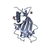
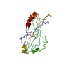
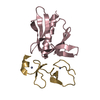
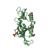
- PDB-1zv9: Crystal structure analysis of a type II cohesin domain from the c... -
+ Open data

- Basic information
| Entry | Database: PDB / ID: 1zv9 | ||||||
|---|---|---|---|---|---|---|---|
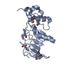
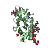
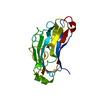
| Title | Crystal structure analysis of a type II cohesin domain from the cellulosome of Acetivibrio cellulolyticus- SeMet derivative | ||||||
 Components Components | Cellulosomal scaffoldin adaptor protein B | ||||||
 Keywords Keywords |  cellulosome / cohesins II cellulosome / cohesins II | ||||||
| Function / homology |  Function and homology information Function and homology informationpolysaccharide catabolic process / hydrolase activity, hydrolyzing O-glycosyl compounds / carbohydrate binding / extracellular region / metal ion bindingSimilarity search - Function | ||||||
| Biological species |  Acetivibrio cellulolyticus (bacteria) Acetivibrio cellulolyticus (bacteria) | ||||||
| Method | X-RAY DIFFRACTION / SYNCHROTRON / MOLECULAR REPLACEMENT / Resolution: 1.28 Å | ||||||
 Authors Authors | Noach, I. / Rosenheck, S. / Lamed, R. / Shimon, L. / Bayer, E. / Frolow, F. | ||||||
 Citation Citation | Journal: J.Mol.Biol. / Year: 2009 Title: Intermodular linker flexibility revealed from crystal structures of adjacent cellulosomal cohesins of Acetivibrio cellulolyticus. Authors: Noach, I. / Frolow, F. / Alber, O. / Lamed, R. / Shimon, L.J. / Bayer, E.A. #1: Journal: Acta Crystallogr.,Sect.D / Year: 2003Title: Preliminary X-ray characterisation and phasing of a type II cohesin domain from the cellulosome of Acetivibrio cellulolyticus Authors: Noach, I. / Lamed, R. / Xu, Q. / Rosenheck, S. / Shimon, L.J.W. / Bayer, E. / Frolow, F. | ||||||
| History |
|
- Structure visualization
| Structure viewer | Molecule: MolmilJmol/JSmol |
|---|
- Downloads & links
-Download
| PDBx/mmCIF format | 1zv9.cif.gz | 98 KB | Display | PDBx/mmCIF format |
|---|---|---|---|---|
| PDB format | pdb1zv9.ent.gz | 73.5 KB | Display | PDB format |
| PDBx/mmJSON format | 1zv9.json.gz | Tree view | PDBx/mmJSON format | |
| Others |  Other downloads Other downloads |
-Validation report
| Arichive directory | https://data.pdbj.org/pub/pdb/validation_reports/zv/1zv9ftp://data.pdbj.org/pub/pdb/validation_reports/zv/1zv9 | HTTPS FTP |
|---|
-Related structure data
| Related structure data |  3bwzC  3fnkC  3ghpC  1qznS S: Starting model for refinement C: citing same article ( |
|---|---|
| Similar structure data |


-Links
 PDBj
PDBj


- Assembly
| Deposited unit | 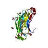
| ||||||||||
|---|---|---|---|---|---|---|---|---|---|---|---|
| 1 |
| ||||||||||
| Unit cell |
|
-Components
-Protein , 1 types, 1 molecules A
| #1: Protein | Mass: 18529.287 Da / Num. of mol.: 1 / Fragment: cohesin II domain from cellulosome assembly / Mutation: MET41MSE,MET63MSE,MET139MSE Source method: isolated from a genetically manipulated source Source: (gene. exp.) Acetivibrio cellulolyticus (bacteria) / Gene: ScaB / Plasmid: pet28a / Species (production host): Escherichia coli / Production host: Escherichia coli BL21 (bacteria) / Strain (production host): BL21 / References: GenBank: 31540575, UniProt: Q7WYN3*PLUS |
|---|
-Non-polymers , 6 types, 307 molecules 

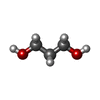


| #2: Chemical | Nitrate Mass: 62.005 Da / Num. of mol.: 2 / Source method: obtained synthetically / Formula: NO3 Mass: 62.005 Da / Num. of mol.: 2 / Source method: obtained synthetically / Formula: NO3#3: Chemical | ChemComp-EDO / Ethylene glycol Mass: 62.068 Da / Num. of mol.: 6 / Source method: obtained synthetically / Formula: C2H6O2 Mass: 62.068 Da / Num. of mol.: 6 / Source method: obtained synthetically / Formula: C2H6O2#4: Chemical | ChemComp-PDO / | 1,3-Propanediol Mass: 76.094 Da / Num. of mol.: 1 / Source method: obtained synthetically / Formula: C3H8O2 Mass: 76.094 Da / Num. of mol.: 1 / Source method: obtained synthetically / Formula: C3H8O2#5: Chemical | ChemComp-FMT / | Formic acid Mass: 46.025 Da / Num. of mol.: 1 / Source method: obtained synthetically / Formula: CH2O2 Mass: 46.025 Da / Num. of mol.: 1 / Source method: obtained synthetically / Formula: CH2O2#6: Chemical | Acetic acid Mass: 60.052 Da / Num. of mol.: 2 / Source method: obtained synthetically / Formula: C2H4O2 Mass: 60.052 Da / Num. of mol.: 2 / Source method: obtained synthetically / Formula: C2H4O2#7: Water | ChemComp-HOH / | WaterMass: 18.015 Da / Num. of mol.: 295 / Source method: isolated from a natural source / Formula: H2O |
|---|
-Experimental details
-Experiment
| Experiment | Method: X-RAY DIFFRACTION / Number of used crystals: 1 |
|---|
- Sample preparation
| Crystal | Density Matthews: 2.5 Å3/Da / Density % sol: 51.4 % |
|---|---|
| Crystal grow | Temperature: 293 K / Method: vapor diffusion / pH: 6 Details: Ammonium sulfate, pH 6, vapor diffusion, temperature 293K |
-Data collection
| Diffraction | Mean temperature: 100 K |
|---|---|
| Diffraction source | Source: SYNCHROTRON / Site: ESRF  / Beamline: ID14-2 / Wavelength: 0.933 Å / Beamline: ID14-2 / Wavelength: 0.933 Å |
| Detector | Type: ADSC QUANTUM 4 / Detector: CCD / Date: Jun 16, 2003 / Details: Mirrors |
| Radiation | Monochromator: Si 111 channel / Protocol: SINGLE WAVELENGTH / Monochromatic (M) / Laue (L): M / Scattering type: x-ray |
| Radiation wavelength | Wavelength: 0.933 Å / Relative weight: 1 |
| Reflection | Resolution: 1.28→50 Å / Num. all: 49349 / Num. obs: 48796 / % possible obs: 98.9 % / Observed criterion σ(I): -3 / Redundancy: 12.8 % / Biso Wilson estimate: 19.2 Å2 / Rmerge(I) obs: 0.065 / Rsym value: 0.065 / Χ2: 1.23 / Net I/σ(I): 24.2 |
| Reflection shell | Resolution: 1.28→1.3 Å / Redundancy: 10.2 % / Rmerge(I) obs: 0.49 / Mean I/σ(I) obs: 2.6 / Num. unique all: 2340 / Rsym value: 0.49 / Χ2: 0.841 / % possible all: 97.1 |
- Processing
| Software |
| ||||||||||||||||||||||||||||||||||||||||||||||||||||||||||||||||||||||||||||||||||||||||||||||||||||||||||||||||||||||||||||||||||||||||||||
|---|---|---|---|---|---|---|---|---|---|---|---|---|---|---|---|---|---|---|---|---|---|---|---|---|---|---|---|---|---|---|---|---|---|---|---|---|---|---|---|---|---|---|---|---|---|---|---|---|---|---|---|---|---|---|---|---|---|---|---|---|---|---|---|---|---|---|---|---|---|---|---|---|---|---|---|---|---|---|---|---|---|---|---|---|---|---|---|---|---|---|---|---|---|---|---|---|---|---|---|---|---|---|---|---|---|---|---|---|---|---|---|---|---|---|---|---|---|---|---|---|---|---|---|---|---|---|---|---|---|---|---|---|---|---|---|---|---|---|---|---|---|
| Refinement | Method to determine structure: MOLECULAR REPLACEMENT Starting model: PDB ENTRY 1QZN Resolution: 1.28→46.63 Å / Cor.coef. Fo:Fc: 0.981 / Cor.coef. Fo:Fc free: 0.974 / SU B: 1.182 / SU ML: 0.023 / SU R Cruickshank DPI: 0.035 / Isotropic thermal model: Anisotropic / Cross valid method: THROUGHOUT / σ(F): 0 / ESU R Free: 0.039 / Stereochemistry target values: Engh & Huber
| ||||||||||||||||||||||||||||||||||||||||||||||||||||||||||||||||||||||||||||||||||||||||||||||||||||||||||||||||||||||||||||||||||||||||||||
| Solvent computation | Ion probe radii: 0.8 Å / Shrinkage radii: 0.8 Å / VDW probe radii: 1.2 Å / Solvent model: BABINET MODEL WITH MASK | ||||||||||||||||||||||||||||||||||||||||||||||||||||||||||||||||||||||||||||||||||||||||||||||||||||||||||||||||||||||||||||||||||||||||||||
| Displacement parameters | Biso mean: 12.949 Å2
| ||||||||||||||||||||||||||||||||||||||||||||||||||||||||||||||||||||||||||||||||||||||||||||||||||||||||||||||||||||||||||||||||||||||||||||
| Refinement step | Cycle: LAST / Resolution: 1.28→46.63 Å
| ||||||||||||||||||||||||||||||||||||||||||||||||||||||||||||||||||||||||||||||||||||||||||||||||||||||||||||||||||||||||||||||||||||||||||||
| Refine LS restraints |
| ||||||||||||||||||||||||||||||||||||||||||||||||||||||||||||||||||||||||||||||||||||||||||||||||||||||||||||||||||||||||||||||||||||||||||||
| LS refinement shell | Resolution: 1.28→1.312 Å / Total num. of bins used: 20
|
