 Movie
Movie Controller


+ Open data

- Basic information
| Entry | Database: PDB / ID: 1ztd | ||||||
|---|---|---|---|---|---|---|---|
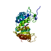
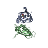
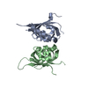
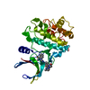
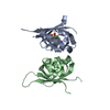
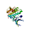
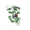
| Title | Hypothetical Protein Pfu-631545-001 From Pyrococcus furiosus | ||||||
 Components Components | Hypothetical Protein Pfu-631545-001 Hypothesis Hypothesis | ||||||
 Keywords Keywords | STRUCTURAL GENOMICS / UNKNOWN FUNCTION / SOUTHEAST COLLABORATORY FOR STRUCTURAL GENOMICS / SECSG / PROTEIN STRUCTURE INITIATIVE / PSI / CONSERVED HYPOTHETICAL PROTEIN / PYROCOCCUS FURIOSUS / HYPERTHERMOPHILE / Pfu-631545-001 | ||||||
| Function / homology |  Function and homology information Function and homology information | ||||||
| Biological species |   Pyrococcus furiosus (archaea) Pyrococcus furiosus (archaea) | ||||||
| Method | X-RAY DIFFRACTION / SYNCHROTRON / SAD / Resolution: 2 Å | ||||||
 Authors Authors | Fu, Z.-Q. / Horanyi, P. / Florence, Q. / Liu, Z.-J. / Chen, L. / Lee, D. / Habel, J. / Xu, H. / Nguyen, D. / Chang, S.-H. ...Fu, Z.-Q. / Horanyi, P. / Florence, Q. / Liu, Z.-J. / Chen, L. / Lee, D. / Habel, J. / Xu, H. / Nguyen, D. / Chang, S.-H. / Zhou, W. / Zhang, H. / Jenney Jr., F.E. / Sha, B. / Adams, M.W.W. / Rose, J.P. / Wang, B.-C. / Southeast Collaboratory for Structural Genomics (SECSG) | ||||||
 Citation Citation | Journal: To be Published Title: Hypothetical Protein Pfu-631545-001 From Pyrococcus furiosus Authors: Fu, Z.-Q. / Horanyi, P. / Florence, Q. / Liu, Z.-J. / Chen, L. / Lee, D. / Habel, J. / Xu, H. / Nguyen, D. / Chang, S.-H. / Zhou, W. / Zhang, H. / Jenney Jr., F.E. / Sha, B. / Adams, M.W.W. ...Authors: Fu, Z.-Q. / Horanyi, P. / Florence, Q. / Liu, Z.-J. / Chen, L. / Lee, D. / Habel, J. / Xu, H. / Nguyen, D. / Chang, S.-H. / Zhou, W. / Zhang, H. / Jenney Jr., F.E. / Sha, B. / Adams, M.W.W. / Rose, J.P. / Wang, B.-C. | ||||||
| History |
| ||||||
| Remark 999 | SEQUENCE Currently there is no protein sequence database reference available for this entry. |
- Structure visualization
| Structure viewer | Molecule: MolmilJmol/JSmol |
|---|
- Downloads & links
-Download
| PDBx/mmCIF format | 1ztd.cif.gz | 59.8 KB | Display | PDBx/mmCIF format |
|---|---|---|---|---|
| PDB format | pdb1ztd.ent.gz | 44 KB | Display | PDB format |
| PDBx/mmJSON format | 1ztd.json.gz | Tree view | PDBx/mmJSON format | |
| Others |  Other downloads Other downloads |
-Validation report
| Arichive directory | https://data.pdbj.org/pub/pdb/validation_reports/zt/1ztdftp://data.pdbj.org/pub/pdb/validation_reports/zt/1ztd | HTTPS FTP |
|---|
-Related structure data
| Similar structure data | |
|---|---|
| Other databases |


-Links
 PDBj
PDBj

- Assembly
| Deposited unit | 
| ||||||||
|---|---|---|---|---|---|---|---|---|---|
| 1 |
| ||||||||
| Unit cell |
|
-Components
| #1: Protein | Hypothesis Mass: 14735.063 Da / Num. of mol.: 2 Source method: isolated from a genetically manipulated source Source: (gene. exp.) Pyrococcus furiosus (archaea) / Plasmid: PET24D BAM / Production host:  Escherichia coli (E. coli) / Strain (production host): C41 PRIL / References: UniProt: Q8U363 Escherichia coli (E. coli) / Strain (production host): C41 PRIL / References: UniProt: Q8U363#2: Water | ChemComp-HOH / | Water Mass: 18.015 Da / Num. of mol.: 63 / Source method: isolated from a natural source / Formula: H2O Mass: 18.015 Da / Num. of mol.: 63 / Source method: isolated from a natural source / Formula: H2O |
|---|
-Experimental details
-Experiment
| Experiment | Method: X-RAY DIFFRACTION / Number of used crystals: 1 |
|---|
- Sample preparation
| Crystal | Density Matthews: 2.06 Å3/Da / Density % sol: 40 % |
|---|---|
| Crystal grow | Temperature: 291 K / pH: 8.2 Details: 5% w/v PEG-8000, pH 8.2, Sitting Drop, Vapor Diffusion, temperature 291K, pH 8.20 |
-Data collection
| Diffraction | Mean temperature: 100 K |
|---|---|
| Diffraction source | Source: SYNCHROTRON / Site: APS  / Beamline: 22-ID / Wavelength: 0.97941 / Beamline: 22-ID / Wavelength: 0.97941 |
| Detector | Type: MARMOSAIC 225 mm CCD / Detector: CCD / Date: Apr 1, 2004 |
| Radiation | Protocol: SINGLE WAVELENGTH / Monochromatic (M) / Laue (L): M / Scattering type: x-ray |
| Radiation wavelength | Wavelength: 0.97941 Å / Relative weight: 1 |
| Reflection | Resolution: 2→35.97 Å / Num. obs: 15035 / % possible obs: 98.5 % / Observed criterion σ(I): 1 / Redundancy: 9.92 % / Biso Wilson estimate: 14.6 Å2 / Rmerge(I) obs: 0.0825 / Rsym value: 0.0825 / Net I/σ(I): 18.72 |
| Reflection shell | Resolution: 2→2.07 Å / Redundancy: 7.6 % / Rmerge(I) obs: 0.229 / Mean I/σ(I) obs: 5.07 / Rsym value: 0.229 / % possible all: 86.3 |
- Processing
| Software |
| ||||||||||||||||||||||||||||||||||||||||||||||||||||||||||||||||||||||||||||||||
|---|---|---|---|---|---|---|---|---|---|---|---|---|---|---|---|---|---|---|---|---|---|---|---|---|---|---|---|---|---|---|---|---|---|---|---|---|---|---|---|---|---|---|---|---|---|---|---|---|---|---|---|---|---|---|---|---|---|---|---|---|---|---|---|---|---|---|---|---|---|---|---|---|---|---|---|---|---|---|---|---|---|
| Refinement | Method to determine structure: SAD / Resolution: 2→35.97 Å / Rfactor Rfree error: 0.01 / Data cutoff high absF: 260232.73 / Data cutoff low absF: 0 / Isotropic thermal model: RESTRAINED / Cross valid method: THROUGHOUT / σ(F): 0 / Stereochemistry target values: ENGH & HUBER
| ||||||||||||||||||||||||||||||||||||||||||||||||||||||||||||||||||||||||||||||||
| Solvent computation | Solvent model: FLAT MODEL / Bsol: 63.89 Å2 / ksol: 0.38 e/Å3 | ||||||||||||||||||||||||||||||||||||||||||||||||||||||||||||||||||||||||||||||||
| Displacement parameters | Biso mean: 25.99 Å2
| ||||||||||||||||||||||||||||||||||||||||||||||||||||||||||||||||||||||||||||||||
| Refine analyze |
| ||||||||||||||||||||||||||||||||||||||||||||||||||||||||||||||||||||||||||||||||
| Refinement step | Cycle: LAST / Resolution: 2→35.97 Å
| ||||||||||||||||||||||||||||||||||||||||||||||||||||||||||||||||||||||||||||||||
| Refine LS restraints |
| ||||||||||||||||||||||||||||||||||||||||||||||||||||||||||||||||||||||||||||||||
| LS refinement shell | Resolution: 2→2.07 Å / Rfactor Rfree error: 0.031 / Total num. of bins used: 10
| ||||||||||||||||||||||||||||||||||||||||||||||||||||||||||||||||||||||||||||||||
| Xplor file |
|
