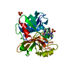
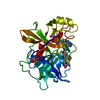
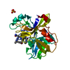
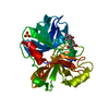
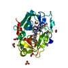
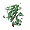
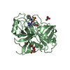
- PDB-1zm8: Apo Crystal structure of Nuclease A from Anabaena sp. -
+
Open data
ID or keywords:
Loading...
-
Basic information
Entry
Database: PDB / ID: 1zm8
Title
Apo Crystal structure of Nuclease A from Anabaena sp.
Components
Nuclease
Keywords
HYDROLASE / NucA / nuclease / metal dependent
Function / homology
Function and homology information
Hydrolases; Acting on ester bonds; Endoribonucleases that are active with either ribo- or deoxyribonucleic acids and produce 5'-phosphomonoesters / endonuclease activity / nucleic acid binding / periplasmic space / metal ion binding Similarity search - Function
DNA/RNA non-specific endonuclease, active site / DNA/RNA non-specific endonucleases active site. / Non-specific endonuclease / Extracellular Endonuclease; Chain A / Extracellular Endonuclease, subunit A / Extracellular Endonuclease, subunit A / DNA/RNA non-specific endonuclease / DNA/RNA non-specific endonuclease / DNA/RNA non-specific endonuclease / DNA/RNA non-specific endonuclease ...DNA/RNA non-specific endonuclease, active site / DNA/RNA non-specific endonucleases active site. / Non-specific endonuclease / Extracellular Endonuclease; Chain A / Extracellular Endonuclease, subunit A / Extracellular Endonuclease, subunit A / DNA/RNA non-specific endonuclease / DNA/RNA non-specific endonuclease / DNA/RNA non-specific endonuclease / DNA/RNA non-specific endonuclease / DNA/RNA non-specific endonuclease superfamily / His-Me finger superfamily / 3-Layer(aba) Sandwich / Alpha Beta Similarity search - Domain/homology
Mass: 28542.887 Da / Num. of mol.: 1 / Fragment: catalytic domain (residues 25-274) / Mutation: D121A Source method: isolated from a genetically manipulated source Source: (gene. exp.) Anabaena sp. (bacteria) / Gene: nucA / Plasmid: PET / Production host: Escherichia coli (E. coli) / Strain (production host): BL21 star (DE3) References: UniProt: P38446, Hydrolases; Acting on ester bonds; Endoribonucleases that are active with either ribo- or deoxyribonucleic acids and produce 5'-phosphomonoesters
In the structure databanks used in Yorodumi, some data are registered as the other names, "COVID-19 virus" and "2019-nCoV". Here are the details of the virus and the list of structure data.
Jan 31, 2019. EMDB accession codes are about to change! (news from PDBe EMDB page)
EMDB accession codes are about to change! (news from PDBe EMDB page)
The allocation of 4 digits for EMDB accession codes will soon come to an end. Whilst these codes will remain in use, new EMDB accession codes will include an additional digit and will expand incrementally as the available range of codes is exhausted. The current 4-digit format prefixed with “EMD-” (i.e. EMD-XXXX) will advance to a 5-digit format (i.e. EMD-XXXXX), and so on. It is currently estimated that the 4-digit codes will be depleted around Spring 2019, at which point the 5-digit format will come into force.
The EM Navigator/Yorodumi systems omit the EMD- prefix.
Related info.:Q: What is EMD? / ID/Accession-code notation in Yorodumi/EM Navigator
Yorodumi is a browser for structure data from EMDB, PDB, SASBDB, etc.
This page is also the successor to EM Navigator detail page, and also detail information page/front-end page for Omokage search.
The word "yorodu" (or yorozu) is an old Japanese word meaning "ten thousand". "mi" (miru) is to see.
Related info.:EMDB / PDB / SASBDB / Comparison of 3 databanks / Yorodumi Search / Aug 31, 2016. New EM Navigator & Yorodumi / Yorodumi Papers / Jmol/JSmol / Function and homology information / Changes in new EM Navigator and Yorodumi
 Movie
Movie Controller


 Open data
 Basic information
 Components
 Keywords
 Function and homology information
 Anabaena sp. (bacteria)
 Authors
 Citation
 Structure visualization
 Downloads & links
 Other downloads
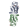


 PDBj
PDBj Assembly
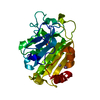

 Mass: 54.938 Da / Num. of mol.: 3 / Source method: obtained synthetically / Formula: Mn
 Mass: 96.063 Da / Num. of mol.: 2 / Source method: obtained synthetically / Formula: SO4
 Mass: 22.990 Da / Num. of mol.: 1 / Source method: obtained synthetically / Formula: Na
 Mass: 18.015 Da / Num. of mol.: 145 / Source method: isolated from a natural source / Formula: H2O
 Sample preparation
 Processing