 Movie
Movie Controller

[English] 日本語
 Yorodumi
- PDB-1zld: Crystal structure of a RGD-containing host-selective toxin: Pyren... -
+ Open data

- Basic information
| Entry | Database: PDB / ID: 1zld | |||||||||
|---|---|---|---|---|---|---|---|---|---|---|
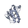
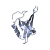
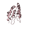
| Title | Crystal structure of a RGD-containing host-selective toxin: Pyrenophora tritici-repentis Ptr ToxA | |||||||||
 Components Components | Ptr necrosis toxin | |||||||||
 Keywords Keywords |  TOXIN / beta-sandwich / RGD-motif TOXIN / beta-sandwich / RGD-motif | |||||||||
| Function / homology | Proteinaceous host-selective toxin ToxA / Proteinaceous host-selective toxin ToxA / ToxA superfamily / Proteinaceous host-selective toxin ToxA / Immunoglobulin-like / Sandwich / Mainly Beta / A necrosis toxin Function and homology information Function and homology information | |||||||||
| Biological species |  Pyrenophora tritici-repentis (fungus) Pyrenophora tritici-repentis (fungus) | |||||||||
| Method | X-RAY DIFFRACTION / SYNCHROTRON / SAD / Resolution: 1.65 Å | |||||||||
 Authors Authors | Sarma, G.N. / Manning, V.A. / Ciuffetti, L.M. / Karplus, P.A. | |||||||||
 Citation Citation | Journal: Plant Cell / Year: 2005 Title: Structure of Ptr ToxA: An RGD-Containing Host-Selective Toxin from Pyrenophora tritici-repentis Authors: Sarma, G.N. / Manning, V.A. / Ciuffetti, L.M. / Karplus, P.A. | |||||||||
| History |
|
- Structure visualization
| Structure viewer | Molecule: MolmilJmol/JSmol |
|---|
- Downloads & links
-Download
| PDBx/mmCIF format | 1zld.cif.gz | 53.7 KB | Display | PDBx/mmCIF format |
|---|---|---|---|---|
| PDB format | pdb1zld.ent.gz | 42.7 KB | Display | PDB format |
| PDBx/mmJSON format | 1zld.json.gz | Tree view | PDBx/mmJSON format | |
| Others |  Other downloads Other downloads |
-Validation report
| Arichive directory | https://data.pdbj.org/pub/pdb/validation_reports/zl/1zldftp://data.pdbj.org/pub/pdb/validation_reports/zl/1zld | HTTPS FTP |
|---|
-Related structure data


-Links
 PDBj
PDBj- Assembly
| Deposited unit | 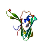
| ||||||||||
|---|---|---|---|---|---|---|---|---|---|---|---|
| 1 |
| ||||||||||
| Unit cell |
| ||||||||||
| Components on special symmetry positions |
|
-Components
| #1: Protein | Mass: 13207.755 Da / Num. of mol.: 1 / Fragment: C-terminal domain / Source method: isolated from a natural source / Source: (natural) Pyrenophora tritici-repentis (fungus) / References: UniProt: P78737 | ||
|---|---|---|---|
| #2: Chemical | Sulfate  Mass: 96.063 Da / Num. of mol.: 2 / Source method: obtained synthetically / Formula: SO4 Mass: 96.063 Da / Num. of mol.: 2 / Source method: obtained synthetically / Formula: SO4#3: Water | ChemComp-HOH / | Water Mass: 18.015 Da / Num. of mol.: 105 / Source method: isolated from a natural source / Formula: H2O Mass: 18.015 Da / Num. of mol.: 105 / Source method: isolated from a natural source / Formula: H2O |
-Experimental details
-Experiment
| Experiment | Method: X-RAY DIFFRACTION / Number of used crystals: 1 |
|---|
- Sample preparation
| Crystal | Density Matthews: 3 Å3/Da / Density % sol: 60 % |
|---|---|
| Crystal grow | Temperature: 300 K / Method: vapor diffusion, hanging drop / pH: 6.5 Details: 0.5 M Ammonium sulfate, 15% Dioxane, 0.1 M MES, pH 6.5, VAPOR DIFFUSION, HANGING DROP, temperature 300K |
-Data collection
| Diffraction | Mean temperature: 100 K |
|---|---|
| Diffraction source | Source: SYNCHROTRON / Site: ALS  / Beamline: 8.2.1 / Wavelength: 1 Å / Beamline: 8.2.1 / Wavelength: 1 Å |
| Detector | Type: ADSC QUANTUM 210 / Detector: CCD / Date: Jan 15, 2004 |
| Radiation | Monochromator: Double crystal, Si(111) / Protocol: SINGLE WAVELENGTH / Monochromatic (M) / Laue (L): M / Scattering type: x-ray |
| Radiation wavelength | Wavelength: 1 Å / Relative weight: 1 |
| Reflection | Resolution: 1.65→50 Å / Num. all: 19466 / Num. obs: 19466 / % possible obs: 100 % / Observed criterion σ(F): 0 / Observed criterion σ(I): 0 / Redundancy: 7.6 % |
| Reflection shell | Resolution: 1.65→1.71 Å / Redundancy: 4.7 % / % possible all: 100 |
- Processing
| Software |
| ||||||||||||||||||||
|---|---|---|---|---|---|---|---|---|---|---|---|---|---|---|---|---|---|---|---|---|---|
| Refinement | Method to determine structure: SAD / Resolution: 1.65→50 Å / σ(F): 0 / Stereochemistry target values: Engh & Huber
| ||||||||||||||||||||
| Refinement step | Cycle: LAST / Resolution: 1.65→50 Å
| ||||||||||||||||||||
| Refine LS restraints |
|
