 Movie
Movie Controller

[English] 日本語
 Yorodumi
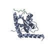
- PDB-1zko: Crystal structure of Glycine cleavage system H protein (tm0212) f... -
+ Open data

- Basic information
| Entry | Database: PDB / ID: 1zko | ||||||
|---|---|---|---|---|---|---|---|
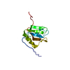
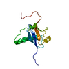
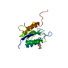
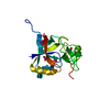
| Title | Crystal structure of Glycine cleavage system H protein (tm0212) from Thermotoga maritima at 1.65 A resolution | ||||||
 Components Components | Glycine cleavage system H protein | ||||||
 Keywords Keywords | Glycine cleavage H-protein / tm0212 / Glycine cleavage system H protein / Structural Genomics / Joint Center for Structural Genomics / JCSG / Protein Structure Initiative / PSI | ||||||
| Function / homology |  Function and homology informationglycine cleavage complex / glycine decarboxylation via glycine cleavage system / cytosol / cytoplasm Function and homology informationglycine cleavage complex / glycine decarboxylation via glycine cleavage system / cytosol / cytoplasmSimilarity search - Function | ||||||
| Biological species |   Thermotoga maritima (bacteria) Thermotoga maritima (bacteria) | ||||||
| Method | X-RAY DIFFRACTION / SYNCHROTRON / MAD / Resolution: 1.65 Å | ||||||
 Authors Authors | Joint Center for Structural Genomics (JCSG) | ||||||
 Citation Citation | Journal: To be published Title: Crystal structure of Glycine cleavage system H protein (tm0212) from Thermotoga maritima at 1.65 A resolution Authors: Joint Center for Structural Genomics (JCSG) | ||||||
| History |
| ||||||
| Remark 999 | SEQUENCE CLONING ARTIFACT: IN THE CLONING CONSTRUCT, THE START CODON WAS CHANGED FROM ATG TO TTG. ...SEQUENCE CLONING ARTIFACT: IN THE CLONING CONSTRUCT, THE START CODON WAS CHANGED FROM ATG TO TTG. AS A RESULT, THE FIRST METHIONINE RESIDUE IS MUTATED TO LEUCINE. |
- Structure visualization
| Structure viewer | Molecule: MolmilJmol/JSmol |
|---|
- Downloads & links
-Download
| PDBx/mmCIF format | 1zko.cif.gz | 67.1 KB | Display | PDBx/mmCIF format |
|---|---|---|---|---|
| PDB format | pdb1zko.ent.gz | 52.2 KB | Display | PDB format |
| PDBx/mmJSON format | 1zko.json.gz | Tree view | PDBx/mmJSON format | |
| Others |  Other downloads Other downloads |
-Validation report
| Arichive directory | https://data.pdbj.org/pub/pdb/validation_reports/zk/1zkoftp://data.pdbj.org/pub/pdb/validation_reports/zk/1zko | HTTPS FTP |
|---|
-Related structure data
| Similar structure data | |
|---|---|
| Other databases |


-Links
 PDBj
PDBj

- Assembly
| Deposited unit | 
| ||||||||||||||||||
|---|---|---|---|---|---|---|---|---|---|---|---|---|---|---|---|---|---|---|---|
| 1 | 
| ||||||||||||||||||
| 2 | 
| ||||||||||||||||||
| 3 | 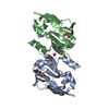
| ||||||||||||||||||
| Unit cell |
| ||||||||||||||||||
| Noncrystallographic symmetry (NCS) | NCS domain:
NCS domain segments: Component-ID: 1 / Ens-ID: 1 / Beg label comp-ID: HIS / End label comp-ID: GLN / Refine code: 6 / Auth seq-ID: -4 - 123 / Label seq-ID: 8 - 135
|
-Components
| #1: Protein | Mass: 15513.833 Da / Num. of mol.: 2 Source method: isolated from a genetically manipulated source Source: (gene. exp.) Thermotoga maritima (bacteria) / Gene: gcvH / Production host: Escherichia coli (E. coli) / References: UniProt: Q9WY55#2: Chemical | ChemComp-NA / |   Mass: 22.990 Da / Num. of mol.: 1 / Source method: obtained synthetically / Formula: Na Mass: 22.990 Da / Num. of mol.: 1 / Source method: obtained synthetically / Formula: Na#3: Water | ChemComp-HOH / | Water Mass: 18.015 Da / Num. of mol.: 249 / Source method: isolated from a natural source / Formula: H2O Mass: 18.015 Da / Num. of mol.: 249 / Source method: isolated from a natural source / Formula: H2O |
|---|
-Experimental details
-Experiment
| Experiment | Method: X-RAY DIFFRACTION / Number of used crystals: 1 |
|---|
- Sample preparation
| Crystal | Density Matthews: 1.87 Å3/Da / Density % sol: 33.63 % |
|---|---|
| Crystal grow | Temperature: 277 K / Method: vapor diffusion, sitting drop, nanodrop / pH: 4 Details: 1.0M LiCl, 30.0% PEG-6000, 0.1M Citrate pH 4.0, VAPOR DIFFUSION,SITTING DROP,NANODROP, temperature 277K |
-Data collection
| Diffraction | Mean temperature: 100 K | |||||||||||||||||||||||||||||||||||||||||||||||||||||||||||||||||||||||||||||
|---|---|---|---|---|---|---|---|---|---|---|---|---|---|---|---|---|---|---|---|---|---|---|---|---|---|---|---|---|---|---|---|---|---|---|---|---|---|---|---|---|---|---|---|---|---|---|---|---|---|---|---|---|---|---|---|---|---|---|---|---|---|---|---|---|---|---|---|---|---|---|---|---|---|---|---|---|---|---|
| Diffraction source | Source: SYNCHROTRON / Site: SSRL  / Beamline: BL11-1 / Wavelength: 0.946415, 0.979129, 0.979494 / Beamline: BL11-1 / Wavelength: 0.946415, 0.979129, 0.979494 | |||||||||||||||||||||||||||||||||||||||||||||||||||||||||||||||||||||||||||||
| Detector | Type: ADSC QUANTUM 315 / Detector: CCD / Date: Apr 4, 2005 / Details: Flat mirror (vertical focusing) | |||||||||||||||||||||||||||||||||||||||||||||||||||||||||||||||||||||||||||||
| Radiation | Monochromator: single crystal Si(111) bent monochromator(horizontal focusing) Protocol: MAD / Monochromatic (M) / Laue (L): M / Scattering type: x-ray | |||||||||||||||||||||||||||||||||||||||||||||||||||||||||||||||||||||||||||||
| Radiation wavelength |
| |||||||||||||||||||||||||||||||||||||||||||||||||||||||||||||||||||||||||||||
| Reflection | Resolution: 1.56→42.53 Å / Num. obs: 30875 / % possible obs: 87.4 % / Redundancy: 3.6 % / Rmerge(I) obs: 0.073 / Rsym value: 0.073 / Net I/σ(I): 6.4 | |||||||||||||||||||||||||||||||||||||||||||||||||||||||||||||||||||||||||||||
| Reflection shell | Diffraction-ID: 1
|
-Phasing
| Phasing | Method: MAD |
|---|
- Processing
| Software |
| ||||||||||||||||||||||||||||||||||||||||||||||||||||||||||||||||||||||||||||||||||||||||||
|---|---|---|---|---|---|---|---|---|---|---|---|---|---|---|---|---|---|---|---|---|---|---|---|---|---|---|---|---|---|---|---|---|---|---|---|---|---|---|---|---|---|---|---|---|---|---|---|---|---|---|---|---|---|---|---|---|---|---|---|---|---|---|---|---|---|---|---|---|---|---|---|---|---|---|---|---|---|---|---|---|---|---|---|---|---|---|---|---|---|---|---|
| Refinement | Method to determine structure: MAD / Resolution: 1.65→42.53 Å / Cor.coef. Fo:Fc: 0.939 / Cor.coef. Fo:Fc free: 0.925 / SU B: 5.538 / SU ML: 0.095 / Cross valid method: THROUGHOUT / ESU R: 0.14 / ESU R Free: 0.135 Stereochemistry target values: MAXIMUM LIKELIHOOD WITH PHASES Details: 1. THE R AND R-FREE FACTORS ARE HIGH DESPITE THE RESOLUTION. ONE REASON MAY BE THAT THE DIFFRACTION IMAGES SHOW DIFFRACTION FROM MORE THAN ONE CRYSTAL. THE ELECTRON DENSITY MAPS ARE VERY ...Details: 1. THE R AND R-FREE FACTORS ARE HIGH DESPITE THE RESOLUTION. ONE REASON MAY BE THAT THE DIFFRACTION IMAGES SHOW DIFFRACTION FROM MORE THAN ONE CRYSTAL. THE ELECTRON DENSITY MAPS ARE VERY CLEAR FOR THE TWO MONOMERS IN THE ASYMMETRIC UNIT.
| ||||||||||||||||||||||||||||||||||||||||||||||||||||||||||||||||||||||||||||||||||||||||||
| Solvent computation | Ion probe radii: 0.8 Å / Shrinkage radii: 0.8 Å / VDW probe radii: 1.2 Å / Solvent model: BABINET MODEL WITH MASK | ||||||||||||||||||||||||||||||||||||||||||||||||||||||||||||||||||||||||||||||||||||||||||
| Displacement parameters | Biso mean: 18.464 Å2
| ||||||||||||||||||||||||||||||||||||||||||||||||||||||||||||||||||||||||||||||||||||||||||
| Refinement step | Cycle: LAST / Resolution: 1.65→42.53 Å
| ||||||||||||||||||||||||||||||||||||||||||||||||||||||||||||||||||||||||||||||||||||||||||
| Refine LS restraints |
| ||||||||||||||||||||||||||||||||||||||||||||||||||||||||||||||||||||||||||||||||||||||||||
| Refine LS restraints NCS | Ens-ID: 1 / Number: 950 / Refine-ID: X-RAY DIFFRACTION
| ||||||||||||||||||||||||||||||||||||||||||||||||||||||||||||||||||||||||||||||||||||||||||
| LS refinement shell | Resolution: 1.651→1.693 Å / Total num. of bins used: 20
|
