 Movie
Movie Controller

[English] 日本語
 Yorodumi
- PDB-1zhn: Crystal Structure of mouse CD1d bound to the self ligand phosphat... -
+ Open data

- Basic information
| Entry | Database: PDB / ID: 1zhn | |||||||||
|---|---|---|---|---|---|---|---|---|---|---|
| Title | Crystal Structure of mouse CD1d bound to the self ligand phosphatidylcholine | |||||||||
 Components Components |
| |||||||||
 Keywords Keywords |  IMMUNE SYSTEM / MEMBRANE PROTEIN / MHC fold / antigen binding domain / Ig fold IMMUNE SYSTEM / MEMBRANE PROTEIN / MHC fold / antigen binding domain / Ig fold | |||||||||
| Function / homology |  Function and homology information Function and homology informationregulation of immature T cell proliferation in thymus / positive regulation of NK T cell differentiation / positive regulation of NK T cell activation / NK T cell differentiation / endogenous lipid antigen binding / exogenous lipid antigen binding / antigen processing and presentation, endogenous lipid antigen via MHC class Ib / antigen processing and presentation, exogenous lipid antigen via MHC class Ib / lipopeptide binding / positive thymic T cell selection ...regulation of immature T cell proliferation in thymus / positive regulation of NK T cell differentiation / positive regulation of NK T cell activation / NK T cell differentiation / endogenous lipid antigen binding / exogenous lipid antigen binding / antigen processing and presentation, endogenous lipid antigen via MHC class Ib / antigen processing and presentation, exogenous lipid antigen via MHC class Ib / lipopeptide binding / positive thymic T cell selection / positive regulation of macrophage activation / Endosomal/Vacuolar pathway / DAP12 interactions / Antigen Presentation: Folding, assembly and peptide loading of class I MHC / ER-Phagosome pathway / DAP12 signaling / Immunoregulatory interactions between a Lymphoid and a non-Lymphoid cell / regulation of membrane depolarization / positive regulation of interleukin-4 production / antigen processing and presentation / regulation of immune response / cellular defense response / T cell receptor binding / positive regulation of interleukin-2 production / Neutrophil degranulation / cellular response to iron(III) ion / antigen processing and presentation of exogenous protein antigen via MHC class Ib, TAP-dependent / negative regulation of forebrain neuron differentiation / regulation of erythrocyte differentiation / peptide antigen assembly with MHC class I protein complex / response to molecule of bacterial origin / regulation of iron ion transport / MHC class I peptide loading complex / HFE-transferrin receptor complex / T cell mediated cytotoxicity / cellular response to iron ion / antigen processing and presentation of endogenous peptide antigen via MHC class I / positive regulation of T cell cytokine production / MHC class I protein complex / multicellular organismal-level iron ion homeostasis / negative regulation of neurogenesis / peptide antigen assembly with MHC class II protein complex / positive regulation of receptor-mediated endocytosis / MHC class II protein complex / cellular response to nicotine / positive regulation of T cell mediated cytotoxicity / phagocytic vesicle membrane / peptide antigen binding / positive regulation of cellular senescence / antigen processing and presentation of exogenous peptide antigen via MHC class II / negative regulation of epithelial cell proliferation / positive regulation of immune response / positive regulation of type II interferon production / antimicrobial humoral immune response mediated by antimicrobial peptide / sensory perception of smell / positive regulation of T cell activation / late endosome / negative regulation of neuron projection development / MHC class II protein complex binding / late endosome membrane / T cell differentiation in thymus / iron ion transport / protein refolding / antibacterial humoral response / protein homotetramerization / intracellular iron ion homeostasis / cellular response to lipopolysaccharide / defense response to Gram-negative bacterium / amyloid fibril formation / lysosome / early endosome / learning or memory / endosome membrane / defense response to Gram-positive bacterium / immune response / lysosomal membrane / external side of plasma membrane / innate immune response / structural molecule activity / Golgi apparatus / protein homodimerization activity / extracellular space / identical protein binding / plasma membrane / cytosolSimilarity search - Function | |||||||||
| Biological species |  Mus musculus (house mouse) Mus musculus (house mouse) | |||||||||
| Method | X-RAY DIFFRACTION / SYNCHROTRON / MOLECULAR REPLACEMENT / Resolution: 2.8 Å | |||||||||
 Authors Authors | Giabbai, B. / Sidobre, S. / Crispin, M.M.D. / Sanchez Ruiz, Y. / Bachi, A. / Kronenberg, M. / Wilson, I.A. / Degano, M. | |||||||||
 Citation Citation | Journal: J.Immunol. / Year: 2005 Title: Crystal structure of mouse CD1d bound to the self ligand phosphatidylcholine: a molecular basis for NKT cell activation Authors: Giabbai, B. / Sidobre, S. / Crispin, M.M.D. / Sanchez Ruiz, Y. / Bachi, A. / Kronenberg, M. / Wilson, I.A. / Degano, M. #1: Journal: Science / Year: 1997Title: Crystal structure of mouse CD1: An MHC-like fold with a large hydrophobic binding groove Authors: Zeng, Z. / Castano, A.R. / Segelke, B.W. / Stura, E.A. / Peterson, P.A. / Wilson, I.A. | |||||||||
| History |
|
- Structure visualization
| Structure viewer | Molecule: MolmilJmol/JSmol |
|---|
- Downloads & links
-Download
| PDBx/mmCIF format | 1zhn.cif.gz | 96.7 KB | Display | PDBx/mmCIF format |
|---|---|---|---|---|
| PDB format | pdb1zhn.ent.gz | 70.8 KB | Display | PDB format |
| PDBx/mmJSON format | 1zhn.json.gz | Tree view | PDBx/mmJSON format | |
| Others |  Other downloads Other downloads |
-Validation report
| Arichive directory | https://data.pdbj.org/pub/pdb/validation_reports/zh/1zhnftp://data.pdbj.org/pub/pdb/validation_reports/zh/1zhn | HTTPS FTP |
|---|
-Related structure data
| Related structure data |  1cd1S S: Starting model for refinement |
|---|---|
| Similar structure data |


-Links
 PDBj
PDBj


- Assembly
| Deposited unit | 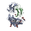
| ||||||||||
|---|---|---|---|---|---|---|---|---|---|---|---|
| 1 |
| ||||||||||
| Unit cell |
| ||||||||||
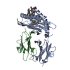
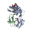
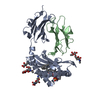
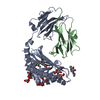
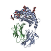
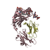
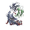
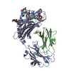
| Details | The biological assembly is a heterodimeric CD1d/beta-2microglobulin complex bound to one PC molecule, as found in the crystal asymmetric unit |
-Components
-Protein , 2 types, 2 molecules AB
| #1: Protein | Mass: 31131.100 Da / Num. of mol.: 1 Source method: isolated from a genetically manipulated source Details: Self phospholipid copurified with protein / Source: (gene. exp.) Mus musculus (house mouse) / Cell line (production host): S2 cells / Production host:  Drosophila melanogaster (fruit fly) / References: GenBank: 12832671, UniProt: P11609*PLUS Drosophila melanogaster (fruit fly) / References: GenBank: 12832671, UniProt: P11609*PLUS |
|---|---|
| #2: Protein | Beta-2 microglobulin Mass: 11704.359 Da / Num. of mol.: 1 Source method: isolated from a genetically manipulated source Source: (gene. exp.) Mus musculus (house mouse) / Cell line (production host): S2 cells / Production host: Drosophila melanogaster (fruit fly) / References: UniProt: P01887 |
-Sugars , 3 types, 3 molecules 
| #3: Polysaccharide | alpha-L-fucopyranose-(1-6)-2-acetamido-2-deoxy-beta-D-glucopyranose / Mass: 367.349 Da / Num. of mol.: 1 Source method: isolated from a genetically manipulated source |
|---|---|
| #4: Polysaccharide | alpha-D-mannopyranose-(1-3)-[alpha-D-mannopyranose-(1-6)]alpha-D-mannopyranose-(1-4)-2-acetamido-2- ...alpha-D-mannopyranose-(1-3)-[alpha-D-mannopyranose-(1-6)]alpha-D-mannopyranose-(1-4)-2-acetamido-2-deoxy-beta-D-glucopyranose-(1-4)-[alpha-L-fucopyranose-(1-6)]2-acetamido-2-deoxy-beta-D-glucopyranose / Mass: 1056.964 Da / Num. of mol.: 1 Source method: isolated from a genetically manipulated source |
| #5: Sugar | ChemComp-NAG / N-Acetylglucosamine Type: D-saccharide, beta linking / Mass: 221.208 Da / Num. of mol.: 1 Type: D-saccharide, beta linking / Mass: 221.208 Da / Num. of mol.: 1Source method: isolated from a genetically manipulated source Formula: C8H15NO6 |
-Non-polymers , 3 types, 52 molecules 


| #6: Chemical | ChemComp-PC6 /  Mass: 804.172 Da / Num. of mol.: 1 / Source method: obtained synthetically / Formula: C45H90NO8P Mass: 804.172 Da / Num. of mol.: 1 / Source method: obtained synthetically / Formula: C45H90NO8P |
|---|---|
| #7: Chemical | ChemComp-GOL / Glycerol Mass: 92.094 Da / Num. of mol.: 1 / Source method: obtained synthetically / Formula: C3H8O3 Mass: 92.094 Da / Num. of mol.: 1 / Source method: obtained synthetically / Formula: C3H8O3 |
| #8: Water | ChemComp-HOH / WaterMass: 18.015 Da / Num. of mol.: 50 / Source method: isolated from a natural source / Formula: H2O |
-Experimental details
-Experiment
| Experiment | Method: X-RAY DIFFRACTION / Number of used crystals: 1 |
|---|
- Sample preparation
| Crystal | Density Matthews: 2.9 Å3/Da / Density % sol: 57.7 % |
|---|---|
| Crystal grow | Temperature: 293 K / Method: vapor diffusion, hanging drop / pH: 7.4 Details: 100 mM Tris, 200 mM NH4(SO4)2, 25% PEG 4000, pH 7.4, VAPOR DIFFUSION, HANGING DROP, temperature 293K |
-Data collection
| Diffraction | Mean temperature: 100 K |
|---|---|
| Diffraction source | Source: SYNCHROTRON / Site: SSRL  / Beamline: BL7-1 / Wavelength: 1 Å / Beamline: BL7-1 / Wavelength: 1 Å |
| Detector | Type: MARRESEARCH / Detector: IMAGE PLATE / Date: May 22, 1998 |
| Radiation | Monochromator: Mirrors / Protocol: SINGLE WAVELENGTH / Monochromatic (M) / Laue (L): M / Scattering type: x-ray |
| Radiation wavelength | Wavelength: 1 Å / Relative weight: 1 |
| Reflection | Resolution: 2.8→76.7 Å / Num. all: 12734 / Num. obs: 12734 / % possible obs: 98.1 % / Observed criterion σ(I): -3 / Redundancy: 4.3 % / Biso Wilson estimate: 39.3 Å2 / Rsym value: 0.056 / Net I/σ(I): 8.4 |
| Reflection shell | Resolution: 2.8→2.95 Å / Redundancy: 4 % / Mean I/σ(I) obs: 2.5 / Num. unique all: 1810 / Rsym value: 0.29 / % possible all: 96 |
- Processing
| Software |
| ||||||||||||||||||||||||||||||||||||||||||||||||||||||||||||||||||||||||||||||||||||||||||||||||||||||||||||||||||||||||||||||||||||||||||||||||||||||||||||||||||||||||||
|---|---|---|---|---|---|---|---|---|---|---|---|---|---|---|---|---|---|---|---|---|---|---|---|---|---|---|---|---|---|---|---|---|---|---|---|---|---|---|---|---|---|---|---|---|---|---|---|---|---|---|---|---|---|---|---|---|---|---|---|---|---|---|---|---|---|---|---|---|---|---|---|---|---|---|---|---|---|---|---|---|---|---|---|---|---|---|---|---|---|---|---|---|---|---|---|---|---|---|---|---|---|---|---|---|---|---|---|---|---|---|---|---|---|---|---|---|---|---|---|---|---|---|---|---|---|---|---|---|---|---|---|---|---|---|---|---|---|---|---|---|---|---|---|---|---|---|---|---|---|---|---|---|---|---|---|---|---|---|---|---|---|---|---|---|---|---|---|---|---|---|---|
| Refinement | Method to determine structure: MOLECULAR REPLACEMENT Starting model: 1CD1 Resolution: 2.8→76.7 Å / Cor.coef. Fo:Fc: 0.896 / Cor.coef. Fo:Fc free: 0.838 / SU B: 16.19 / SU ML: 0.309 / Cross valid method: THROUGHOUT / σ(F): 0 / ESU R Free: 0.43 / Stereochemistry target values: MAXIMUM LIKELIHOOD / Details: HYDROGENS HAVE BEEN ADDED IN THE RIDING POSITIONS
| ||||||||||||||||||||||||||||||||||||||||||||||||||||||||||||||||||||||||||||||||||||||||||||||||||||||||||||||||||||||||||||||||||||||||||||||||||||||||||||||||||||||||||
| Solvent computation | Ion probe radii: 0.8 Å / Shrinkage radii: 0.8 Å / VDW probe radii: 1.2 Å / Solvent model: BABINET MODEL WITH MASK | ||||||||||||||||||||||||||||||||||||||||||||||||||||||||||||||||||||||||||||||||||||||||||||||||||||||||||||||||||||||||||||||||||||||||||||||||||||||||||||||||||||||||||
| Displacement parameters | Biso mean: 25.724 Å2
| ||||||||||||||||||||||||||||||||||||||||||||||||||||||||||||||||||||||||||||||||||||||||||||||||||||||||||||||||||||||||||||||||||||||||||||||||||||||||||||||||||||||||||
| Refinement step | Cycle: LAST / Resolution: 2.8→76.7 Å
| ||||||||||||||||||||||||||||||||||||||||||||||||||||||||||||||||||||||||||||||||||||||||||||||||||||||||||||||||||||||||||||||||||||||||||||||||||||||||||||||||||||||||||
| Refine LS restraints |
| ||||||||||||||||||||||||||||||||||||||||||||||||||||||||||||||||||||||||||||||||||||||||||||||||||||||||||||||||||||||||||||||||||||||||||||||||||||||||||||||||||||||||||
| LS refinement shell | Resolution: 2.8→2.873 Å / Total num. of bins used: 20
|
