 Movie
Movie Controller

[English] 日本語
 Yorodumi
- PDB-1zh2: Crystal Structure Of The Calcium-Bound Receiver Domain Of Kdp Pot... -
+ Open data

- Basic information
| Entry | Database: PDB / ID: 1zh2 | ||||||
|---|---|---|---|---|---|---|---|
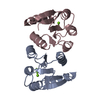
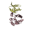
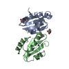
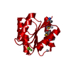
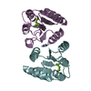
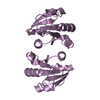
| Title | Crystal Structure Of The Calcium-Bound Receiver Domain Of Kdp Potassium Transport System Response Regulator KdpE | ||||||
 Components Components | KDP operon transcriptional regulatory protein kdpE | ||||||
 Keywords Keywords |  TRANSCRIPTION / Two-Component System / Gene Regulation / Transcription Factor / Kdp Potassium Transport System / Doubly Wound Five-Stranded Beta-Alpha Fold TRANSCRIPTION / Two-Component System / Gene Regulation / Transcription Factor / Kdp Potassium Transport System / Doubly Wound Five-Stranded Beta-Alpha Fold | ||||||
| Function / homology |  Function and homology information Function and homology informationphosphorelay response regulator activity / DNA-binding transcription activator activity / cis-regulatory region sequence-specific DNA binding / protein-DNA complex / transcription cis-regulatory region binding / DNA-binding transcription factor activity / regulation of DNA-templated transcription / protein homodimerization activity / cytosolSimilarity search - Function | ||||||
| Biological species |  Escherichia coli (E. coli) Escherichia coli (E. coli) | ||||||
| Method | X-RAY DIFFRACTION / SYNCHROTRON / MOLECULAR REPLACEMENT / Resolution: 2 Å | ||||||
 Authors Authors | Toro-Roman, A. / Wu, T. / Stock, A.M. | ||||||
 Citation Citation | Journal: Protein Sci. / Year: 2005 Title: A common dimerization interface in bacterial response regulators KdpE and TorR. Authors: Toro-Roman, A. / Wu, T. / Stock, A.M. | ||||||
| History |
| ||||||
| Remark 700 | SHEET Determination method: author |
- Structure visualization
| Structure viewer | Molecule: MolmilJmol/JSmol |
|---|
- Downloads & links
-Download
| PDBx/mmCIF format | 1zh2.cif.gz | 62.8 KB | Display | PDBx/mmCIF format |
|---|---|---|---|---|
| PDB format | pdb1zh2.ent.gz | 45.5 KB | Display | PDB format |
| PDBx/mmJSON format | 1zh2.json.gz | Tree view | PDBx/mmJSON format | |
| Others |  Other downloads Other downloads |
-Validation report
| Arichive directory | https://data.pdbj.org/pub/pdb/validation_reports/zh/1zh2ftp://data.pdbj.org/pub/pdb/validation_reports/zh/1zh2 | HTTPS FTP |
|---|
-Related structure data
| Related structure data | 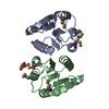 1zgzC 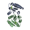 1zh4C  1xhfS  1zh3 C: citing same article ( S: Starting model for refinement |
|---|---|
| Similar structure data |


-Links
 PDBj
PDBj

- Assembly
| Deposited unit | 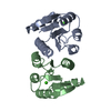
| |||||||||
|---|---|---|---|---|---|---|---|---|---|---|
| 1 |
| |||||||||
| Unit cell |
| |||||||||
| Components on special symmetry positions |
|
-Components
| #1: Protein | Mass: 13488.371 Da / Num. of mol.: 2 / Fragment: N-Terminal Receiver Domain (residues 1-121) / Mutation: A121Q Source method: isolated from a genetically manipulated source Source: (gene. exp.) Escherichia coli (E. coli) / Gene: kdpE / Plasmid: pEF27 (pJES307 derivative) / Species (production host): Escherichia coli / Production host: Escherichia coli BL21(DE3) (bacteria) / Strain (production host): BL21(DE3) / References: UniProt: P21866#2: Chemical |   Mass: 40.078 Da / Num. of mol.: 2 / Source method: obtained synthetically / Formula: Ca Mass: 40.078 Da / Num. of mol.: 2 / Source method: obtained synthetically / Formula: Ca#3: Water | ChemComp-HOH / | Water Mass: 18.015 Da / Num. of mol.: 158 / Source method: isolated from a natural source / Formula: H2O Mass: 18.015 Da / Num. of mol.: 158 / Source method: isolated from a natural source / Formula: H2O |
|---|
-Experimental details
-Experiment
| Experiment | Method: X-RAY DIFFRACTION / Number of used crystals: 1 |
|---|
- Sample preparation
| Crystal | Density Matthews: 2.7 Å3/Da / Density % sol: 54.2 % |
|---|---|
| Crystal grow | Temperature: 293 K / Method: vapor diffusion, hanging drop / pH: 6 Details: PEG 8000, Calcium Acetate, MES Buffer, pH 6.0, VAPOR DIFFUSION, HANGING DROP, temperature 293K |
-Data collection
| Diffraction | Mean temperature: 100 K |
|---|---|
| Diffraction source | Source: SYNCHROTRON / Site: NSLS  / Beamline: X4A / Wavelength: 1.07179 Å / Beamline: X4A / Wavelength: 1.07179 Å |
| Detector | Type: ADSC QUANTUM 4 / Detector: CCD / Date: Nov 11, 2004 |
| Radiation | Monochromator: KOHZU Double Crystal Monochromator With A Sagittally Focused Second Crystal. Crystal Type Is Si(111) Protocol: SINGLE WAVELENGTH / Monochromatic (M) / Laue (L): M / Scattering type: x-ray |
| Radiation wavelength | Wavelength: 1.07179 Å / Relative weight: 1 |
| Reflection | Resolution: 2→30 Å / Num. all: 20179 / Num. obs: 20163 / % possible obs: 100 % / Observed criterion σ(I): -3 / Redundancy: 13.6 % / Biso Wilson estimate: 12.3 Å2 / Rsym value: 0.063 / Net I/σ(I): 48 |
| Reflection shell | Resolution: 2→2.07 Å / Redundancy: 10.2 % / Mean I/σ(I) obs: 24.2 / Num. unique all: 1992 / Rsym value: 0.159 / % possible all: 99.9 |
- Processing
| Software |
| |||||||||||||||||||||||||||||||||||||||||||||||||||||||||||||||||||||||||||
|---|---|---|---|---|---|---|---|---|---|---|---|---|---|---|---|---|---|---|---|---|---|---|---|---|---|---|---|---|---|---|---|---|---|---|---|---|---|---|---|---|---|---|---|---|---|---|---|---|---|---|---|---|---|---|---|---|---|---|---|---|---|---|---|---|---|---|---|---|---|---|---|---|---|---|---|---|
| Refinement | Method to determine structure: MOLECULAR REPLACEMENT Starting model: PDB ENTRY 1XHF (Ala Model) Resolution: 2→30 Å / Cor.coef. Fo:Fc: 0.941 / Cor.coef. Fo:Fc free: 0.914 / SU B: 3.629 / SU ML: 0.104 / Cross valid method: THROUGHOUT / σ(F): 0 / ESU R: 0.181 / ESU R Free: 0.168 / Stereochemistry target values: MAXIMUM LIKELIHOOD
| |||||||||||||||||||||||||||||||||||||||||||||||||||||||||||||||||||||||||||
| Solvent computation | Ion probe radii: 0.8 Å / Shrinkage radii: 0.8 Å / VDW probe radii: 1.4 Å / Solvent model: BABINET MODEL WITH MASK | |||||||||||||||||||||||||||||||||||||||||||||||||||||||||||||||||||||||||||
| Displacement parameters | Biso mean: 25.051 Å2
| |||||||||||||||||||||||||||||||||||||||||||||||||||||||||||||||||||||||||||
| Refinement step | Cycle: LAST / Resolution: 2→30 Å
| |||||||||||||||||||||||||||||||||||||||||||||||||||||||||||||||||||||||||||
| Refine LS restraints |
| |||||||||||||||||||||||||||||||||||||||||||||||||||||||||||||||||||||||||||
| LS refinement shell | Resolution: 2.001→2.052 Å / Total num. of bins used: 20
|
