 Movie
Movie Controller

[English] 日本語
 Yorodumi
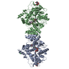
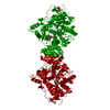
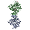
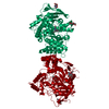
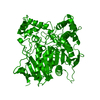
- PDB-1zgc: Crystal Structure of Torpedo Californica Acetylcholinesterase in ... -
+ Open data

- Basic information
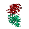
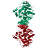
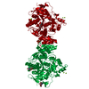
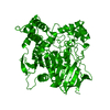
| Entry | Database: PDB / ID: 1zgc | ||||||
|---|---|---|---|---|---|---|---|
| Title | Crystal Structure of Torpedo Californica Acetylcholinesterase in Complex With an (RS)-Tacrine(10)-Hupyridone Inhibitor. | ||||||
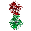
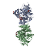
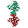
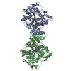
 Components Components | Acetylcholinesterase | ||||||
 Keywords Keywords | HYDROLASE / SERINE-HYDROLASE / PROTEIN-INHIBITOR COMPLEX / ENANTIOMERIC SELECTIVITY / Israel Structural Proteomics Center / ISPC / Structural Genomics | ||||||
| Function / homology |  Function and homology information Function and homology informationacetylcholine catabolic process in synaptic cleft / acetylcholinesterase / acetylcholinesterase activity / choline metabolic process / side of membrane / synaptic cleft / synapse / extracellular space / plasma membraneSimilarity search - Function | ||||||
| Biological species |   Torpedo californica (Pacific electric ray) Torpedo californica (Pacific electric ray) | ||||||
| Method | X-RAY DIFFRACTION / MOLECULAR REPLACEMENT / Resolution: 2.1 Å | ||||||
 Authors Authors | Haviv, H. / Wong, D.M. / Greenblatt, H.M. / Carlier, P.R. / Pang, Y.P. / Silman, I. / Sussman, J.L. / Israel Structural Proteomics Center (ISPC) | ||||||
 Citation Citation | Journal: J.Am.Chem.Soc. / Year: 2005 Title: Crystal Packing Mediates Enantioselective Ligand Recognition at the Peripheral Site of Acetylcholinesterase Authors: Haviv, H. / Wong, D.M. / Greenblatt, H.M. / Carlier, P.R. / Pang, Y.P. / Silman, I. / Sussman, J.L. #1: Journal: BIOORG.MED.CHEM.LETT. / Year: 1999 Title: Potent, easily synthesized huperzine A-tacrine hybrid acetylcholinesterase inhibitors Authors: Carlier, P.R. / Du, D.-M. / Han, Y.-F. / Liu, J. / Pang, Y.-P. #2: Journal: J.Am.Chem.Soc. / Year: 2003Title: Acetylcholinesterase Complexed with Bivalent Ligands Related to Huperzine A: Experimental Evidence for Species-Dependent Protein-Ligand Complementarity Authors: Wong, D.M. / Greenblatt, H.M. / Dvir, H. / Carlier, P.R. / Han, Y.-F. / Pang, Y.-P. / Silman, I. / Sussman, J.L. #3: Journal: J.Am.Chem.Soc. / Year: 2004Title: The complex of a bivalent derivative of galanthamine with torpedo acetylcholinesterase displays drastic deformation of the active-site gorge: implications for structure-based drug design Authors: Greenblatt, H.M. / Guillou, C. / Guenard, D. / Argaman, A. / Botti, S. / Badet, B. / Thal, C. / Silman, I. / Sussman, J.L. #4: Journal: Science / Year: 1991 Title: Atomic structure of acetylcholinesterase from Torpedo californica: a prototypic acetylcholine-binding protein Authors: Sussman, J.L. / Harel, M. / Frolow, F. / Oefner, C. / Goldman, A. / Toker, L. / Silman, I. | ||||||
| History |
|
- Structure visualization
| Structure viewer | Molecule: MolmilJmol/JSmol |
|---|
- Downloads & links
-Download
| PDBx/mmCIF format | 1zgc.cif.gz | 223.4 KB | Display | PDBx/mmCIF format |
|---|---|---|---|---|
| PDB format | pdb1zgc.ent.gz | 183 KB | Display | PDB format |
| PDBx/mmJSON format | 1zgc.json.gz | Tree view | PDBx/mmJSON format | |
| Others |  Other downloads Other downloads |
-Validation report
| Arichive directory | https://data.pdbj.org/pub/pdb/validation_reports/zg/1zgcftp://data.pdbj.org/pub/pdb/validation_reports/zg/1zgc | HTTPS FTP |
|---|
-Related structure data

-Links
 PDBj
PDBj
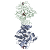
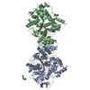
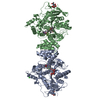
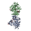
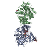
- Assembly
| Deposited unit | 
| ||||||||
|---|---|---|---|---|---|---|---|---|---|
| 1 |
| ||||||||
| Unit cell |
|
-Components
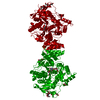
| #1: Protein | / AChE Mass: 61325.090 Da / Num. of mol.: 2 / Source method: isolated from a natural source Source: (natural) Torpedo californica (Pacific electric ray)References: UniProt: P04058, acetylcholinesterase#2: Sugar | ChemComp-NAG / N-Acetylglucosamine  Type: D-saccharide, beta linking / Mass: 221.208 Da / Num. of mol.: 5 / Source method: obtained synthetically / Formula: C8H15NO6 Type: D-saccharide, beta linking / Mass: 221.208 Da / Num. of mol.: 5 / Source method: obtained synthetically / Formula: C8H15NO6#3: Chemical |   Mass: 500.718 Da / Num. of mol.: 2 / Source method: obtained synthetically / Formula: C32H44N4O Mass: 500.718 Da / Num. of mol.: 2 / Source method: obtained synthetically / Formula: C32H44N4O#4: Water | ChemComp-HOH / | Water Mass: 18.015 Da / Num. of mol.: 341 / Source method: isolated from a natural source / Formula: H2O Mass: 18.015 Da / Num. of mol.: 341 / Source method: isolated from a natural source / Formula: H2O |
|---|
-Experimental details
-Experiment
| Experiment | Method: X-RAY DIFFRACTION / Number of used crystals: 1 |
|---|
- Sample preparation
| Crystal | Density Matthews: 2.99 Å3/Da / Density % sol: 58.84 % |
|---|---|
| Crystal grow | Temperature: 277 K Details: PEG 200, pH 5.8, temperature 277K, VAPOR DIFFUSION, SITTING DROP. PROTEIN WAS CRYSTALLISED FROM 28-30% V/V PEG 200 0.5 M MES PH 5.8 AT 277K, WITHOUT SEEDING WITH MICROCRYSTALS; THEN SOAKED ...Details: PEG 200, pH 5.8, temperature 277K, VAPOR DIFFUSION, SITTING DROP. PROTEIN WAS CRYSTALLISED FROM 28-30% V/V PEG 200 0.5 M MES PH 5.8 AT 277K, WITHOUT SEEDING WITH MICROCRYSTALS; THEN SOAKED IN MOTHER LIQUOR (40% V/V PEG 200 IN 0.1 M MES BUFFER, PH 5.8) CONTAINING 4MM (RS)-(+/-)- TACRINE(10)-HUPYRIDONE ((5RS)-(+/-)-5-{[10-(1,2,3,4- TETRAHYDROACRIDIN-9-YLAMINO)DECYL]AMINO}5,6,7,8-TETRAHYDRO-QUINOLIN-2(1H)-ONE) BIS-OXALATE FOR 40 HOURS. |
-Data collection
| Diffraction | Mean temperature: 120 K |
|---|---|
| Diffraction source | Source: ROTATING ANODE / Type: RIGAKU RUH3R / Wavelength: 1.5418 |
| Detector | Type: RIGAKU RAXIS IV / Detector: IMAGE PLATE / Date: Oct 2, 2003 / Details: OSMIC BLUE CONFOCAL MIRRORS |
| Radiation | Protocol: SINGLE WAVELENGTH / Monochromatic (M) / Laue (L): M / Scattering type: x-ray |
| Radiation wavelength | Wavelength: 1.5418 Å / Relative weight: 1 |
| Reflection | Resolution: 2.1→34.5 Å / Num. obs: 84805 / % possible obs: 99.4 % / Redundancy: 0.61 % / Rmerge(I) obs: 0.078 / Net I/σ(I): 18.1 |
| Reflection shell | Resolution: 2.1→2.16 Å / Redundancy: 9.8 % / Rmerge(I) obs: 0.396 / Mean I/σ(I) obs: 3.83 / % possible all: 98.5 |
- Processing
| Software |
| ||||||||||||||||||||||||||||||||||||||||||||||||||||||||||||||||||||||||||||||||||||||||||||||||||||||||||||||||||||||||||||||||||||||||||||||||||||||||||||||||||||||||||
|---|---|---|---|---|---|---|---|---|---|---|---|---|---|---|---|---|---|---|---|---|---|---|---|---|---|---|---|---|---|---|---|---|---|---|---|---|---|---|---|---|---|---|---|---|---|---|---|---|---|---|---|---|---|---|---|---|---|---|---|---|---|---|---|---|---|---|---|---|---|---|---|---|---|---|---|---|---|---|---|---|---|---|---|---|---|---|---|---|---|---|---|---|---|---|---|---|---|---|---|---|---|---|---|---|---|---|---|---|---|---|---|---|---|---|---|---|---|---|---|---|---|---|---|---|---|---|---|---|---|---|---|---|---|---|---|---|---|---|---|---|---|---|---|---|---|---|---|---|---|---|---|---|---|---|---|---|---|---|---|---|---|---|---|---|---|---|---|---|---|---|---|
| Refinement | Method to determine structure: MOLECULAR REPLACEMENT / Resolution: 2.1→34.5 Å / Cor.coef. Fo:Fc: 0.952 / Cor.coef. Fo:Fc free: 0.925 / SU B: 4.485 / SU ML: 0.117 / Cross valid method: THROUGHOUT / ESU R: 0.187 / ESU R Free: 0.176 / Stereochemistry target values: MAXIMUM LIKELIHOOD / Details: HYDROGENS HAVE BEEN ADDED IN THE RIDING POSITIONS
| ||||||||||||||||||||||||||||||||||||||||||||||||||||||||||||||||||||||||||||||||||||||||||||||||||||||||||||||||||||||||||||||||||||||||||||||||||||||||||||||||||||||||||
| Solvent computation | Ion probe radii: 0.8 Å / Shrinkage radii: 0.8 Å / VDW probe radii: 1.4 Å / Solvent model: BABINET MODEL WITH MASK | ||||||||||||||||||||||||||||||||||||||||||||||||||||||||||||||||||||||||||||||||||||||||||||||||||||||||||||||||||||||||||||||||||||||||||||||||||||||||||||||||||||||||||
| Displacement parameters | Biso mean: 24.2 Å2
| ||||||||||||||||||||||||||||||||||||||||||||||||||||||||||||||||||||||||||||||||||||||||||||||||||||||||||||||||||||||||||||||||||||||||||||||||||||||||||||||||||||||||||
| Refinement step | Cycle: LAST / Resolution: 2.1→34.5 Å
| ||||||||||||||||||||||||||||||||||||||||||||||||||||||||||||||||||||||||||||||||||||||||||||||||||||||||||||||||||||||||||||||||||||||||||||||||||||||||||||||||||||||||||
| Refine LS restraints |
| ||||||||||||||||||||||||||||||||||||||||||||||||||||||||||||||||||||||||||||||||||||||||||||||||||||||||||||||||||||||||||||||||||||||||||||||||||||||||||||||||||||||||||
| LS refinement shell | Resolution: 2.1→2.16 Å / Total num. of bins used: 20 /
|
