 Movie
Movie Controller

[English] 日本語
 Yorodumi
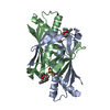
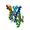
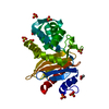
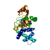
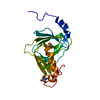
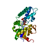
- PDB-1zb9: Crystal structure of Xylella fastidiosa organic peroxide resistan... -
+ Open data

- Basic information
| Entry | Database: PDB / ID: 1zb9 | ||||||
|---|---|---|---|---|---|---|---|
| Title | Crystal structure of Xylella fastidiosa organic peroxide resistance protein | ||||||
 Components Components | organic hydroperoxide resistance protein | ||||||
 Keywords Keywords |  OXIDOREDUCTASE OXIDOREDUCTASE | ||||||
| Function / homology |  Function and homology information Function and homology information | ||||||
| Biological species |  Xylella fastidiosa (bacteria) Xylella fastidiosa (bacteria) | ||||||
| Method | X-RAY DIFFRACTION / SYNCHROTRON / MOLECULAR REPLACEMENT / Resolution: 1.8 Å | ||||||
 Authors Authors | Oliveira, M.A. / Guimaraes, B.G. / Cussiol, J.R. / Medrano, F.J. / Vidigal, S.A. / Gozzo, F.C. / Netto, L.E. | ||||||
 Citation Citation | Journal: J.Mol.Biol. / Year: 2006 Title: Structural Insights into Enzyme-Substrate Interaction and Characterization of Enzymatic Intermediates of Organic Hydroperoxide Resistance Protein from Xylella fastidiosa. Authors: Oliveira, M.A. / Guimaraes, B.G. / Cussiol, J.R. / Medrano, F.J. / Gozzo, F.C. / Netto, L.E. | ||||||
| History |
|
- Structure visualization
| Structure viewer | Molecule: MolmilJmol/JSmol |
|---|
- Downloads & links
-Download
| PDBx/mmCIF format | 1zb9.cif.gz | 65.6 KB | Display | PDBx/mmCIF format |
|---|---|---|---|---|
| PDB format | pdb1zb9.ent.gz | 53 KB | Display | PDB format |
| PDBx/mmJSON format | 1zb9.json.gz | Tree view | PDBx/mmJSON format | |
| Others |  Other downloads Other downloads |
-Validation report
| Arichive directory | https://data.pdbj.org/pub/pdb/validation_reports/zb/1zb9ftp://data.pdbj.org/pub/pdb/validation_reports/zb/1zb9 | HTTPS FTP |
|---|
-Related structure data


-Links
 PDBj
PDBj- Assembly
| Deposited unit | 
| ||||||||
|---|---|---|---|---|---|---|---|---|---|
| 1 |
| ||||||||
| Unit cell |
| ||||||||
| Details | The biologial assembly is the asymmetric unit (dimer). |
-Components
| #1: Protein | Mass: 14961.105 Da / Num. of mol.: 2 Source method: isolated from a genetically manipulated source Source: (gene. exp.) Xylella fastidiosa (bacteria) / Strain: 9a5c / Gene: ohr / Plasmid: pET15b / Species (production host): Escherichia coli / Production host: Escherichia coli BL21 (bacteria) / Strain (production host): BL21 / References: UniProt: Q9PCF4#2: Chemical | Polyethylene glycol  Mass: 354.436 Da / Num. of mol.: 2 / Source method: obtained synthetically / Formula: C16H34O8 / Comment: precipitant*YM Mass: 354.436 Da / Num. of mol.: 2 / Source method: obtained synthetically / Formula: C16H34O8 / Comment: precipitant*YM#3: Water | ChemComp-HOH / | Water Mass: 18.015 Da / Num. of mol.: 256 / Source method: isolated from a natural source / Formula: H2O Mass: 18.015 Da / Num. of mol.: 256 / Source method: isolated from a natural source / Formula: H2O |
|---|
-Experimental details
-Experiment
| Experiment | Method: X-RAY DIFFRACTION / Number of used crystals: 1 |
|---|
- Sample preparation
| Crystal | Density Matthews: 2.8 Å3/Da / Density % sol: 55.4 % |
|---|---|
| Crystal grow | Temperature: 293 K / Method: vapor diffusion, hanging drop / pH: 8.7 Details: Peg 4000, Tris-HCl, 5mM t-BOOH, pH 8.7, VAPOR DIFFUSION, HANGING DROP, temperature 293K |
-Data collection
| Diffraction | Mean temperature: 110 K |
|---|---|
| Diffraction source | Source: SYNCHROTRON / Site: LNLS  / Beamline: D03B-MX1 / Wavelength: 1.453 Å / Beamline: D03B-MX1 / Wavelength: 1.453 Å |
| Detector | Type: MARRESEARCH / Detector: CCD / Date: Oct 5, 2003 / Details: mirrors |
| Radiation | Monochromator: Si 111 / Protocol: SINGLE WAVELENGTH / Monochromatic (M) / Laue (L): M / Scattering type: x-ray |
| Radiation wavelength | Wavelength: 1.453 Å / Relative weight: 1 |
| Reflection | Resolution: 1.8→1.88 Å / Num. obs: 34330 / % possible obs: 99.9 % / Redundancy: 10.8 % / Rsym value: 0.074 / Net I/σ(I): 6.2 |
| Reflection shell | Resolution: 1.8→1.9 Å / Redundancy: 11.1 % / Mean I/σ(I) obs: 2.2 / Num. unique all: 4391 / Rsym value: 0.328 / % possible all: 100 |
- Processing
| Software |
| ||||||||||||||||||||||||||||||||||||||||||||||||||||||||||||||||||||||||||||||||
|---|---|---|---|---|---|---|---|---|---|---|---|---|---|---|---|---|---|---|---|---|---|---|---|---|---|---|---|---|---|---|---|---|---|---|---|---|---|---|---|---|---|---|---|---|---|---|---|---|---|---|---|---|---|---|---|---|---|---|---|---|---|---|---|---|---|---|---|---|---|---|---|---|---|---|---|---|---|---|---|---|---|
| Refinement | Method to determine structure: MOLECULAR REPLACEMENT / Resolution: 1.8→1.88 Å / Stereochemistry target values: Engh & Huber
| ||||||||||||||||||||||||||||||||||||||||||||||||||||||||||||||||||||||||||||||||
| Refinement step | Cycle: LAST / Resolution: 1.8→1.88 Å
| ||||||||||||||||||||||||||||||||||||||||||||||||||||||||||||||||||||||||||||||||
| Refine LS restraints |
| ||||||||||||||||||||||||||||||||||||||||||||||||||||||||||||||||||||||||||||||||
| LS refinement shell | Resolution: 1.8→1.88 Å
| ||||||||||||||||||||||||||||||||||||||||||||||||||||||||||||||||||||||||||||||||
| Xplor file |
|
