 Movie
Movie Controller

[English] 日本語
 Yorodumi
- PDB-1z8k: X-ray structure of allene oxide cyclase from Arabidopsis thaliana... -
+ Open data

- Basic information
| Entry | Database: PDB / ID: 1z8k | ||||||
|---|---|---|---|---|---|---|---|
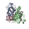
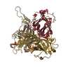
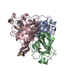
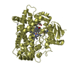
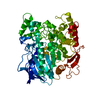
| Title | X-ray structure of allene oxide cyclase from Arabidopsis thaliana at3g25770 | ||||||
 Components Components | At3g25770 protein | ||||||
 Keywords Keywords |  ISOMERASE / STRUCTURAL GENOMICS / PROTEIN STRUCTURE INITIATIVE / PSI / CESG / AT3G25770 / Center for Eukaryotic Structural Genomics / jasmonic acid biosynthesis / plant protein ISOMERASE / STRUCTURAL GENOMICS / PROTEIN STRUCTURE INITIATIVE / PSI / CESG / AT3G25770 / Center for Eukaryotic Structural Genomics / jasmonic acid biosynthesis / plant protein | ||||||
| Function / homology |  Function and homology informationallene-oxide cyclase / allene-oxide cyclase activity / stromule / jasmonic acid biosynthetic process / chloroplast envelope / chloroplast stroma / chloroplast thylakoid membrane / response to cold / chloroplast / cytosol Function and homology informationallene-oxide cyclase / allene-oxide cyclase activity / stromule / jasmonic acid biosynthetic process / chloroplast envelope / chloroplast stroma / chloroplast thylakoid membrane / response to cold / chloroplast / cytosolSimilarity search - Function | ||||||
| Biological species |  Arabidopsis thaliana (thale cress) Arabidopsis thaliana (thale cress) | ||||||
| Method | X-RAY DIFFRACTION / SYNCHROTRON / MAD / Resolution: 1.712 Å | ||||||
 Authors Authors | Wesenberg, G.E. / Phillips Jr., G.N. / Bitto, E. / Bingman, C.A. / Allard, S.T.M. / Center for Eukaryotic Structural Genomics (CESG) | ||||||
 Citation Citation | Journal: To be published Title: X-ray structure of allene oxide cyclase from Arabidopsis thaliana at3g25770 Authors: Center for Eukaryotic Structural Genomics (CESG) | ||||||
| History |
|
- Structure visualization
| Structure viewer | Molecule: MolmilJmol/JSmol |
|---|
- Downloads & links
-Download
| PDBx/mmCIF format | 1z8k.cif.gz | 127.5 KB | Display | PDBx/mmCIF format |
|---|---|---|---|---|
| PDB format | pdb1z8k.ent.gz | 102.8 KB | Display | PDB format |
| PDBx/mmJSON format | 1z8k.json.gz | Tree view | PDBx/mmJSON format | |
| Others |  Other downloads Other downloads |
-Validation report
| Arichive directory | https://data.pdbj.org/pub/pdb/validation_reports/z8/1z8kftp://data.pdbj.org/pub/pdb/validation_reports/z8/1z8k | HTTPS FTP |
|---|
-Related structure data
| Similar structure data | |
|---|---|
| Other databases |

-Links
 PDBj
PDBj- Assembly
| Deposited unit | 
| ||||||||
|---|---|---|---|---|---|---|---|---|---|
| 1 |
| ||||||||
| Unit cell |
|
-Components
| #1: Protein | Mass: 21299.947 Da / Num. of mol.: 3 Source method: isolated from a genetically manipulated source Source: (gene. exp.) Arabidopsis thaliana (thale cress) / Gene: AT3G25770 / Plasmid: PVP-27 / Production host:  Escherichia coli (E. coli) / Strain (production host): B834 P(RARE2) / References: UniProt: Q9LS02, allene-oxide cyclase Escherichia coli (E. coli) / Strain (production host): B834 P(RARE2) / References: UniProt: Q9LS02, allene-oxide cyclase#2: Water | ChemComp-HOH / | Water Mass: 18.015 Da / Num. of mol.: 741 / Source method: isolated from a natural source / Formula: H2O Mass: 18.015 Da / Num. of mol.: 741 / Source method: isolated from a natural source / Formula: H2O |
|---|
-Experimental details
-Experiment
| Experiment | Method: X-RAY DIFFRACTION / Number of used crystals: 1 |
|---|
- Sample preparation
| Crystal | Density Matthews: 2.9 Å3/Da / Density % sol: 57.9 % |
|---|---|
| Crystal grow | Temperature: 293 K / Method: vapor diffusion, hanging drop / pH: 4.5 Details: 10 mg/ml protein, 11% PEG 8K, 0.100 M SODIUM ACETATE, pH 4.5, vapor diffusion, hanging drop, temperature 293K |
-Data collection
| Diffraction |
| |||||||||||||||||||||||||||||||||||||||||||||||||||||||||||||||||||||||||||||||||||||||||||||||||||||||||||||||||||||||||||||||||||||||||||||||||||||||||||||||||||||||||||||||||||||||||||||||||||||||||||||||||||||||||
|---|---|---|---|---|---|---|---|---|---|---|---|---|---|---|---|---|---|---|---|---|---|---|---|---|---|---|---|---|---|---|---|---|---|---|---|---|---|---|---|---|---|---|---|---|---|---|---|---|---|---|---|---|---|---|---|---|---|---|---|---|---|---|---|---|---|---|---|---|---|---|---|---|---|---|---|---|---|---|---|---|---|---|---|---|---|---|---|---|---|---|---|---|---|---|---|---|---|---|---|---|---|---|---|---|---|---|---|---|---|---|---|---|---|---|---|---|---|---|---|---|---|---|---|---|---|---|---|---|---|---|---|---|---|---|---|---|---|---|---|---|---|---|---|---|---|---|---|---|---|---|---|---|---|---|---|---|---|---|---|---|---|---|---|---|---|---|---|---|---|---|---|---|---|---|---|---|---|---|---|---|---|---|---|---|---|---|---|---|---|---|---|---|---|---|---|---|---|---|---|---|---|---|---|---|---|---|---|---|---|---|---|---|---|---|---|---|---|---|
| Diffraction source | Source: SYNCHROTRON / Site: APS  / Beamline: 22-ID / Wavelength: 0.97924, 0.97156, 0.97947 / Beamline: 22-ID / Wavelength: 0.97924, 0.97156, 0.97947 | |||||||||||||||||||||||||||||||||||||||||||||||||||||||||||||||||||||||||||||||||||||||||||||||||||||||||||||||||||||||||||||||||||||||||||||||||||||||||||||||||||||||||||||||||||||||||||||||||||||||||||||||||||||||||
| Detector | Type: MARMOSAIC 300 mm CCD / Detector: CCD / Date: Mar 12, 2005 Details: HORIZONTAL SAGITALLY FOCUSING 2ND BENT MONOCHROMATOR CRYSTAL, VERTICAL BENT FOCUSING MIRROR | |||||||||||||||||||||||||||||||||||||||||||||||||||||||||||||||||||||||||||||||||||||||||||||||||||||||||||||||||||||||||||||||||||||||||||||||||||||||||||||||||||||||||||||||||||||||||||||||||||||||||||||||||||||||||
| Radiation | Monochromator: CRYOGENICALLY COOLED SI (220) DOUBLE BOUNCE / Protocol: MAD / Monochromatic (M) / Laue (L): M / Scattering type: x-ray | |||||||||||||||||||||||||||||||||||||||||||||||||||||||||||||||||||||||||||||||||||||||||||||||||||||||||||||||||||||||||||||||||||||||||||||||||||||||||||||||||||||||||||||||||||||||||||||||||||||||||||||||||||||||||
| Radiation wavelength |
| |||||||||||||||||||||||||||||||||||||||||||||||||||||||||||||||||||||||||||||||||||||||||||||||||||||||||||||||||||||||||||||||||||||||||||||||||||||||||||||||||||||||||||||||||||||||||||||||||||||||||||||||||||||||||
| Reflection | D res low: 50 Å
| |||||||||||||||||||||||||||||||||||||||||||||||||||||||||||||||||||||||||||||||||||||||||||||||||||||||||||||||||||||||||||||||||||||||||||||||||||||||||||||||||||||||||||||||||||||||||||||||||||||||||||||||||||||||||
| Diffraction reflection shell |
| |||||||||||||||||||||||||||||||||||||||||||||||||||||||||||||||||||||||||||||||||||||||||||||||||||||||||||||||||||||||||||||||||||||||||||||||||||||||||||||||||||||||||||||||||||||||||||||||||||||||||||||||||||||||||
| Reflection | Resolution: 1.71→48.166 Å / Num. obs: 73976 / % possible obs: 100 % / Redundancy: 14.1 % / Rmerge(I) obs: 0.075 / Χ2: 1.013 / Net I/σ(I): 19.995 | |||||||||||||||||||||||||||||||||||||||||||||||||||||||||||||||||||||||||||||||||||||||||||||||||||||||||||||||||||||||||||||||||||||||||||||||||||||||||||||||||||||||||||||||||||||||||||||||||||||||||||||||||||||||||
| Reflection shell | Diffraction-ID: 1 / % possible obs: 100 %
|
-Phasing
| Phasing | Method: MAD | |||||||||||||||||||||||||||||||||||||||||||||||||
|---|---|---|---|---|---|---|---|---|---|---|---|---|---|---|---|---|---|---|---|---|---|---|---|---|---|---|---|---|---|---|---|---|---|---|---|---|---|---|---|---|---|---|---|---|---|---|---|---|---|---|
| Phasing set |
| |||||||||||||||||||||||||||||||||||||||||||||||||
| Phasing MAD | D res high: 1.8 Å / D res low: 20 Å / FOM : 0.44 / Reflection: 63255 | |||||||||||||||||||||||||||||||||||||||||||||||||
| Phasing MAD set |
| |||||||||||||||||||||||||||||||||||||||||||||||||
| Phasing MAD set site |
| |||||||||||||||||||||||||||||||||||||||||||||||||
| Phasing MAD shell |
| |||||||||||||||||||||||||||||||||||||||||||||||||
| Phasing dm | FOM : 0.69 / FOM acentric: 0.69 / FOM centric: 0.7 / Reflection: 63255 / Reflection acentric: 57863 / Reflection centric: 5392 | |||||||||||||||||||||||||||||||||||||||||||||||||
| Phasing dm shell |
|
- Processing
| Software |
| |||||||||||||||||||||||||||||||||||||||||||||||||||||||||||||||||||||||||||||||||||||||||||||||||||||||||||||||||||||||||||||||||||||||||||||||||||
|---|---|---|---|---|---|---|---|---|---|---|---|---|---|---|---|---|---|---|---|---|---|---|---|---|---|---|---|---|---|---|---|---|---|---|---|---|---|---|---|---|---|---|---|---|---|---|---|---|---|---|---|---|---|---|---|---|---|---|---|---|---|---|---|---|---|---|---|---|---|---|---|---|---|---|---|---|---|---|---|---|---|---|---|---|---|---|---|---|---|---|---|---|---|---|---|---|---|---|---|---|---|---|---|---|---|---|---|---|---|---|---|---|---|---|---|---|---|---|---|---|---|---|---|---|---|---|---|---|---|---|---|---|---|---|---|---|---|---|---|---|---|---|---|---|---|---|---|---|
| Refinement | Method to determine structure: MAD / Resolution: 1.712→50 Å / Cor.coef. Fo:Fc: 0.965 / Cor.coef. Fo:Fc free: 0.952 / SU B: 1.419 / SU ML: 0.048 / Cross valid method: THROUGHOUT / ESU R: 0.08 / ESU R Free: 0.083 / Stereochemistry target values: MAXIMUM LIKELIHOOD Details: INITIAL MODEL GENERATED BY ARP/WARP, SELENIUM C COEFFICIENT FOR STRUCTURE FACTOR CALCULATION SET TO -9.0000, MOLPROBITY USED TO ASSIST IN FINAL MODEL BUILDING.
| |||||||||||||||||||||||||||||||||||||||||||||||||||||||||||||||||||||||||||||||||||||||||||||||||||||||||||||||||||||||||||||||||||||||||||||||||||
| Solvent computation | Ion probe radii: 0.8 Å / Shrinkage radii: 0.8 Å / VDW probe radii: 1.2 Å / Solvent model: BABINET MODEL WITH MASK | |||||||||||||||||||||||||||||||||||||||||||||||||||||||||||||||||||||||||||||||||||||||||||||||||||||||||||||||||||||||||||||||||||||||||||||||||||
| Displacement parameters | Biso mean: 14.97 Å2
| |||||||||||||||||||||||||||||||||||||||||||||||||||||||||||||||||||||||||||||||||||||||||||||||||||||||||||||||||||||||||||||||||||||||||||||||||||
| Refinement step | Cycle: LAST / Resolution: 1.712→50 Å
| |||||||||||||||||||||||||||||||||||||||||||||||||||||||||||||||||||||||||||||||||||||||||||||||||||||||||||||||||||||||||||||||||||||||||||||||||||
| Refine LS restraints |
| |||||||||||||||||||||||||||||||||||||||||||||||||||||||||||||||||||||||||||||||||||||||||||||||||||||||||||||||||||||||||||||||||||||||||||||||||||
| LS refinement shell | Refine-ID: X-RAY DIFFRACTION / Total num. of bins used: 20
|
