 Movie
Movie Controller


+ Open data

- Basic information
| Entry | Database: PDB / ID: 1z6o | ||||||
|---|---|---|---|---|---|---|---|
| Title | Crystal Structure of Trichoplusia ni secreted ferritin | ||||||
 Components Components |
| ||||||
 Keywords Keywords | METAL BINDING PROTEIN / Iron storage | ||||||
| Function / homology |  Function and homology information Function and homology information ferroxidase / ferroxidase activity / ferric iron binding / iron ion transport / intracellular iron ion homeostasis ferroxidase / ferroxidase activity / ferric iron binding / iron ion transport / intracellular iron ion homeostasisSimilarity search - Function | ||||||
| Biological species |  Trichoplusia ni (cabbage looper) Trichoplusia ni (cabbage looper) | ||||||
| Method | X-RAY DIFFRACTION / SYNCHROTRON / MOLECULAR REPLACEMENT / molecular replacement / Resolution: 1.91 Å | ||||||
 Authors Authors | Hamburger, A.E. / West Jr., A.P. / Hamburger, Z.A. / Hamburger, P. / Bjorkman, P.J. | ||||||
 Citation Citation | Journal: J.Mol.Biol. / Year: 2005 Title: Crystal structure of a secreted insect ferritin reveals a symmetrical arrangement of heavy and light chains. Authors: Hamburger, A.E. / West, A.P. / Hamburger, Z.A. / Hamburger, P. / Bjorkman, P.J. | ||||||
| History |
| ||||||
| Remark 295 | NON-CRYSTALLOGRAPHIC SYMMETRY THE TRANSFORMATIONS PRESENTED ON THE MTRIX RECORDS BELOW DESCRIBE ... NON-CRYSTALLOGRAPHIC SYMMETRY THE TRANSFORMATIONS PRESENTED ON THE MTRIX RECORDS BELOW DESCRIBE NON-CRYSTALLOGRAPHIC RELATIONSHIPS AMONG ATOMS IN THIS ENTRY. APPLYING THE APPROPRIATE MTRIX TRANSFORMATION TO THE RESIDUES LISTED FIRST WILL YIELD APPROXIMATE COORDINATES FOR THE RESIDUES LISTED SECOND. CHAIN IDENTIFIERS GIVEN AS "?" REFER TO CHAINS FOR WHICH ATOMS ARE NOT FOUND IN THIS ENTRY. APPLIED TO TRANSFORMED TO TRANSFORM CHAIN RESIDUES CHAIN RESIDUES RMSD SSS M 1 A 1 .. 212 A 1 .. 212 0.000 M 1 M 1 .. 191 M 1 .. 191 0.000 M 2 A 1 .. 212 B 1 .. 212 0.000 M 2 M 1 .. 191 N 1 .. 191 0.000 M 3 A 1 .. 212 C 1 .. 212 0.000 M 3 M 1 .. 191 O 1 .. 191 0.000 M 4 A 1 .. 212 D 1 .. 212 0.000 M 4 M 1 .. 191 P 1 .. 191 0.000 M 5 A 1 .. 212 E 1 .. 212 0.000 M 5 M 1 .. 191 Q 1 .. 191 0.000 M 6 A 1 .. 212 F 1 .. 212 0.000 M 6 M 1 .. 191 R 1 .. 191 0.000 M 7 A 1 .. 212 G 1 .. 212 0.000 M 7 M 1 .. 191 S 1 .. 191 0.000 M 8 A 1 .. 212 H 1 .. 212 0.000 M 8 M 1 .. 191 T 1 .. 191 0.000 M 9 A 1 .. 212 I 1 .. 212 0.000 M 9 M 1 .. 191 U 1 .. 191 0.000 M 10 A 1 .. 212 J 1 .. 212 0.000 M 10 M 1 .. 191 V 1 .. 191 0.000 M 11 A 1 .. 212 K 1 .. 212 0.000 M 11 M 1 .. 191 W 1 .. 191 0.000 M 12 A 1 .. 212 L 1 .. 212 0.000 M 12 M 1 .. 191 X 1 .. 191 0.000 WHERE SSS -> COLUMNS 8-10 OF MTRIX RECORDS REMARK:NULL | ||||||
| Remark 999 | SEQUENCE GENBANK ENTRIES AAX94728 AND AAX94729 ARE PARTIAL SEQUENCES. THE HEAVY AND LIGHTCHAIN ...SEQUENCE GENBANK ENTRIES AAX94728 AND AAX94729 ARE PARTIAL SEQUENCES. THE HEAVY AND LIGHTCHAIN SEQUENCES IN THIS ENTRY ARE THE COMPLETE AND NATIVE PROCESSED FORMS OF THESE PROTEINS. |
- Structure visualization
| Structure viewer | Molecule: MolmilJmol/JSmol |
|---|
- Downloads & links
-Download
| PDBx/mmCIF format | 1z6o.cif.gz | 1 MB | Display | PDBx/mmCIF format |
|---|---|---|---|---|
| PDB format | pdb1z6o.ent.gz | 856.1 KB | Display | PDB format |
| PDBx/mmJSON format | 1z6o.json.gz | Tree view | PDBx/mmJSON format | |
| Others |  Other downloads Other downloads |
-Validation report
| Arichive directory | https://data.pdbj.org/pub/pdb/validation_reports/z6/1z6oftp://data.pdbj.org/pub/pdb/validation_reports/z6/1z6o | HTTPS FTP |
|---|
-Related structure data
| Related structure data | 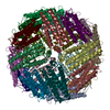 1mfrS S: Starting model for refinement |
|---|---|
| Similar structure data |

-Links
 PDBj
PDBj


- Assembly
| Deposited unit | 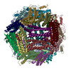
| ||||||||||||||||||||||||||||||||||||||||||||||||||||
|---|---|---|---|---|---|---|---|---|---|---|---|---|---|---|---|---|---|---|---|---|---|---|---|---|---|---|---|---|---|---|---|---|---|---|---|---|---|---|---|---|---|---|---|---|---|---|---|---|---|---|---|---|---|
| 1 |
| ||||||||||||||||||||||||||||||||||||||||||||||||||||
| Unit cell |
| ||||||||||||||||||||||||||||||||||||||||||||||||||||
| Components on special symmetry positions |
| ||||||||||||||||||||||||||||||||||||||||||||||||||||
| Noncrystallographic symmetry (NCS) | NCS oper:
| ||||||||||||||||||||||||||||||||||||||||||||||||||||
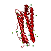
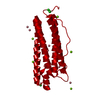
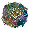
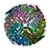
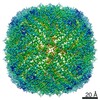
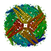
| Details | the asymmetric unit contains one biological assembly composed of 12 heavy and 12 light chains. |

-Components
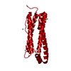
| #1: Protein | Mass: 24341.078 Da / Num. of mol.: 12 / Source method: isolated from a natural source / Source: (natural) Trichoplusia ni (cabbage looper) / Cell line: BTI-Tn-5B1-4 / References: UniProt: Q52SA8#2: Protein | Mass: 21800.779 Da / Num. of mol.: 12 / Source method: isolated from a natural source / Source: (natural) Trichoplusia ni (cabbage looper) / Cell line: BTI-Tn-5B1-4 / References: UniProt: Q52SA9#3: Chemical | ChemComp-FE / Iron  Mass: 55.845 Da / Num. of mol.: 16 / Source method: obtained synthetically / Formula: Fe Mass: 55.845 Da / Num. of mol.: 16 / Source method: obtained synthetically / Formula: Fe#4: Chemical | ChemComp-CA /   Mass: 40.078 Da / Num. of mol.: 8 / Source method: obtained synthetically / Formula: Ca Mass: 40.078 Da / Num. of mol.: 8 / Source method: obtained synthetically / Formula: Ca#5: Water | ChemComp-HOH / | Water Mass: 18.015 Da / Num. of mol.: 4456 / Source method: isolated from a natural source / Formula: H2O Mass: 18.015 Da / Num. of mol.: 4456 / Source method: isolated from a natural source / Formula: H2O |
|---|
-Experimental details
-Experiment
| Experiment | Method: X-RAY DIFFRACTION / Number of used crystals: 1 |
|---|
- Sample preparation
| Crystal | Density Matthews: 2.84 Å3/Da / Density % sol: 56.4 % |
|---|---|
| Crystal grow | Temperature: 277 K / pH: 8 Details: 20 mM Tris, 150 mM NaCl, 0.05% sodium azide, pH 8, spontaneous in storage buffer, temperature 277K, pH 8.00 |
-Data collection
| Diffraction | Mean temperature: 123 K |
|---|---|
| Diffraction source | Source: SYNCHROTRON / Site: ALS  / Beamline: 8.2.2 / Wavelength: 1.0781 / Beamline: 8.2.2 / Wavelength: 1.0781 |
| Detector | Type: ADSC QUANTUM 4 / Detector: CCD / Date: Apr 3, 2004 |
| Radiation | Protocol: SINGLE WAVELENGTH / Monochromatic (M) / Laue (L): M / Scattering type: x-ray |
| Radiation wavelength | Wavelength: 1.0781 Å / Relative weight: 1 |
| Reflection | Resolution: 1.91→29.85 Å / Num. obs: 463420 / % possible obs: 97 % / Redundancy: 1.89 % / Biso Wilson estimate: 18.8 Å2 / Rmerge(I) obs: 0.065 / Net I/σ(I): 11.7 |
| Reflection shell | Resolution: 1.9→1.97 Å / Rmerge(I) obs: 0.344 / Mean I/σ(I) obs: 2.2 / % possible all: 98.7 |
-Phasing
| Phasing | Method: molecular replacement |
|---|
- Processing
| Software |
| ||||||||||||||||||||||||||||||||||||||||||||||||||||||||||||||||||||||||||||||||
|---|---|---|---|---|---|---|---|---|---|---|---|---|---|---|---|---|---|---|---|---|---|---|---|---|---|---|---|---|---|---|---|---|---|---|---|---|---|---|---|---|---|---|---|---|---|---|---|---|---|---|---|---|---|---|---|---|---|---|---|---|---|---|---|---|---|---|---|---|---|---|---|---|---|---|---|---|---|---|---|---|---|
| Refinement | Method to determine structure: MOLECULAR REPLACEMENT Starting model: 1MFR Resolution: 1.91→19.98 Å / Rfactor Rfree error: 0.001 / Occupancy max: 1 / Occupancy min: 0.26 / Cross valid method: THROUGHOUT / Stereochemistry target values: ENGH & HUBER Details: STRICT NCS WAS USED, REFINEMENT WITH NCS CONSTRAINTS WAS PERFORMED USING CHAINS A AND M. THREE IONS(FE3 A 301, CA 5302, CA 5303) AND SIX WATER MOLECULES (HOH 1 TO HOH 6) ARE LOCATED AT ...Details: STRICT NCS WAS USED, REFINEMENT WITH NCS CONSTRAINTS WAS PERFORMED USING CHAINS A AND M. THREE IONS(FE3 A 301, CA 5302, CA 5303) AND SIX WATER MOLECULES (HOH 1 TO HOH 6) ARE LOCATED AT SPECIAL POSITIONS WITH RESPECT TO THE NCS OPERATORS. DURING REFINEMENT THE OCCUPANCIES OF THESE ATOMS WERE 0.26, 0.33, 0.33, 0.33, 0.33, 0.33, 0.33, 0.5, AND 0.5, RESPECTIVELY. IN THIS ENTRY THE OCCUPANCIES OF THESE ATOMS, AND NCS-RELATED ATOMS, ARE TWO OR THREE TIMES HIGHER REFLECTING THE EFFECT OF THE NCS.
| ||||||||||||||||||||||||||||||||||||||||||||||||||||||||||||||||||||||||||||||||
| Solvent computation | Solvent model: CNS BULK SOLVENT MODEL USED / Bsol: 49.6 Å2 / ksol: 0.37 e/Å3 | ||||||||||||||||||||||||||||||||||||||||||||||||||||||||||||||||||||||||||||||||
| Displacement parameters | Biso mean: 28.9 Å2
| ||||||||||||||||||||||||||||||||||||||||||||||||||||||||||||||||||||||||||||||||
| Refine Biso |
| ||||||||||||||||||||||||||||||||||||||||||||||||||||||||||||||||||||||||||||||||
| Refine analyze |
| ||||||||||||||||||||||||||||||||||||||||||||||||||||||||||||||||||||||||||||||||
| Refinement step | Cycle: LAST / Resolution: 1.91→19.98 Å
| ||||||||||||||||||||||||||||||||||||||||||||||||||||||||||||||||||||||||||||||||
| Refine LS restraints |
| ||||||||||||||||||||||||||||||||||||||||||||||||||||||||||||||||||||||||||||||||
| Refine LS restraints NCS | NCS model details: STRICT | ||||||||||||||||||||||||||||||||||||||||||||||||||||||||||||||||||||||||||||||||
| LS refinement shell | Resolution: 1.91→1.97 Å / Rfactor Rfree error: 0.006 / Total num. of bins used: 10
|
