 Movie
Movie Controller


+ Open data

- Basic information
| Entry | Database: PDB / ID: 1yzb | ||||||
|---|---|---|---|---|---|---|---|
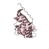
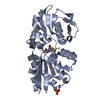
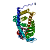
| Title | Solution structure of the Josephin domain of Ataxin-3 | ||||||
 Components Components | Machado-Joseph disease protein 1 Machado–Joseph disease Machado–Joseph disease | ||||||
 Keywords Keywords | TRANSCRIPTION / papain-like fold | ||||||
| Function / homology |  Function and homology information Function and homology informationprotein localization to cytosolic proteasome complex / regulation of cell-substrate adhesion / positive regulation of ERAD pathway / monoubiquitinated protein deubiquitination / intermediate filament cytoskeleton organization / protein K48-linked deubiquitination / nuclear inclusion body / positive regulation of ubiquitin-dependent protein catabolic process / protein K63-linked deubiquitination / cellular response to misfolded protein ...protein localization to cytosolic proteasome complex / regulation of cell-substrate adhesion / positive regulation of ERAD pathway / monoubiquitinated protein deubiquitination / intermediate filament cytoskeleton organization / protein K48-linked deubiquitination / nuclear inclusion body / positive regulation of ubiquitin-dependent protein catabolic process / protein K63-linked deubiquitination / cellular response to misfolded protein / K48-linked deubiquitinase activity / K63-linked deubiquitinase activity / protein quality control for misfolded or incompletely synthesized proteins / FOXO-mediated transcription of oxidative stress, metabolic and neuronal genes / negative regulation of TORC1 signaling / cellular response to amino acid starvation / Josephin domain DUBs / nucleotide-excision repair / mitochondrial membrane / nuclear matrix / microtubule cytoskeleton organization / nervous system development / cellular response to heat / ATPase binding / actin cytoskeleton organization / ubiquitin-dependent protein catabolic process / chemical synaptic transmission / proteasome-mediated ubiquitin-dependent protein catabolic process / ubiquitinyl hydrolase 1 / cysteine-type deubiquitinase activity / mitochondrial matrix / lysosomal membrane / synapse / ubiquitin protein ligase binding / nucleolus / nucleoplasm / identical protein binding / nucleus / plasma membrane / cytosol / cytoplasmSimilarity search - Function | ||||||
| Biological species |  Homo sapiens (human) Homo sapiens (human) | ||||||
| Method | SOLUTION NMR / simulated annealing, torsion angle dynamics | ||||||
 Authors Authors | Nicastro, G. / Masino, L. / Menon, R.P. / Knowles, P.P. / McDonald, N.Q. / Pastore, A. | ||||||
 Citation Citation | Journal: Proc.Natl.Acad.Sci.Usa / Year: 2005 Title: The solution structure of the Josephin domain of ataxin-3: Structural determinants for molecular recognition Authors: Nicastro, G. / Menon, R.P. / Masino, L. / Knowles, P.P. / McDonald, N.Q. / Pastore, A. #1: Journal: J.Biomol.Nmr / Year: 2004 Title: Assignment of the 1H, 13C, and 15N resonances of the Josephin domain of human ataxin-3 Authors: Nicastro, G. / Masino, L. / Frenkiel, T.A. / Kelly, G. / McCormick, J. / Menon, R.P. / Pastore, A. #2: Journal: J.Mol.Biol. / Year: 2004 Title: Characterization of the structure and the amyloidogenic properties of the Josephin domain of the polyglutamine-containing protein ataxin-3 Authors: Masino, L. / Nicastro, G. / Menon, R.P. / Dal Piaz, F. / Calder, L. / Pastore, A. #3: Journal: Febs Lett. / Year: 2003 Title: Domain architecture of the polyglutamine protein ataxin-3: a globular domain followed by a flexible tail Authors: Masino, L. / Musi, V. / Menon, R.P. / Fusi, P. / Kelly, G. / Frenkiel, T.A. / Trottier, Y. / Pastore, A. | ||||||
| History |
|
- Structure visualization
| Structure viewer | Molecule: MolmilJmol/JSmol |
|---|
- Downloads & links
-Download
| PDBx/mmCIF format | 1yzb.cif.gz | 568.3 KB | Display | PDBx/mmCIF format |
|---|---|---|---|---|
| PDB format | pdb1yzb.ent.gz | 473.4 KB | Display | PDB format |
| PDBx/mmJSON format | 1yzb.json.gz | Tree view | PDBx/mmJSON format | |
| Others |  Other downloads Other downloads |
-Validation report
| Arichive directory | https://data.pdbj.org/pub/pdb/validation_reports/yz/1yzbftp://data.pdbj.org/pub/pdb/validation_reports/yz/1yzb | HTTPS FTP |
|---|
-Related structure data
| Similar structure data |
|---|


-Links
 PDBj
PDBj


- Assembly
| Deposited unit | 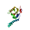
| |||||||||
|---|---|---|---|---|---|---|---|---|---|---|
| 1 |
| |||||||||
| NMR ensembles |
|
-Components
| #1: Protein | Machado–Joseph disease / Ataxin-3 / Spinocerebellar ataxia type 3 protein Mass: 21053.768 Da / Num. of mol.: 1 / Fragment: N-terminal domain of Ataxin-3 Source method: isolated from a genetically manipulated source Source: (gene. exp.) Homo sapiens (human) / Species (production host): Escherichia coli / Production host:  Escherichia coli BL21(DE3) (bacteria) / Strain (production host): BL21(DE3) / References: UniProt: P54252 Escherichia coli BL21(DE3) (bacteria) / Strain (production host): BL21(DE3) / References: UniProt: P54252 |
|---|
-Experimental details
-Experiment
| Experiment | Method: SOLUTION NMR | ||||||||||||
|---|---|---|---|---|---|---|---|---|---|---|---|---|---|
| NMR experiment |
| ||||||||||||
| NMR details | Text: The structure was determined using triple-resonance NMR spectroscopy |
- Sample preparation
| Details | Contents: 0.4mM of Josephin 15N, 13C; 20mM sodium phosphate buffer (pH 6.5); 90% H2O, 10% D2O Solvent system: 90% H2O/10% D2O |
|---|---|
| Sample conditions | Ionic strength: 20mM sodium phosphate / pH: 6.5 / Pressure: ambient / Temperature: 298 K |
-NMR measurement
| NMR spectrometer |
|
|---|
- Processing
| NMR software |
| ||||||||||||||||
|---|---|---|---|---|---|---|---|---|---|---|---|---|---|---|---|---|---|
| Refinement | Method: simulated annealing, torsion angle dynamics / Software ordinal: 1 Details: the structures are based on a total of 5532 unambiguous and 927 ambiguous restraints, 114 dihedral angle restraints, 44 distance restraints from hydrogen bonds. | ||||||||||||||||
| NMR representative | Selection criteria: lowest energy | ||||||||||||||||
| NMR ensemble | Conformer selection criteria: structures with the lowest energy Conformers calculated total number: 100 / Conformers submitted total number: 10 |
