 Movie
Movie Controller

[English] 日本語
 Yorodumi
- PDB-1yxj: Crystal structure of human lectin-like oxidized low-density lipop... -
+ Open data

- Basic information
| Entry | Database: PDB / ID: 1yxj | ||||||
|---|---|---|---|---|---|---|---|
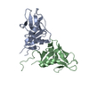
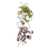
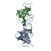
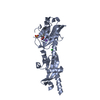
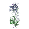
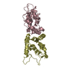
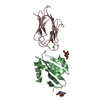
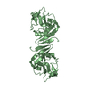
| Title | Crystal structure of human lectin-like oxidized low-density lipoprotein receptor 1 (LOX-1) at low pH | ||||||
 Components Components | oxidised low density lipoprotein (lectin-like) receptor 1 | ||||||
 Keywords Keywords | LIPID BINDING PROTEIN / C-type lectin-like domain /  LOX-1 / CTLD / Scavenger receptor / Oxidized LDL receptor / NK cell receptor LOX-1 / CTLD / Scavenger receptor / Oxidized LDL receptor / NK cell receptor | ||||||
| Function / homology |  Function and homology information Function and homology informationlow-density lipoprotein particle receptor activity / lipoprotein metabolic process / blood circulation / leukocyte cell-cell adhesion / immune system process / tertiary granule membrane / specific granule membrane / Cell surface interactions at the vascular wall / carbohydrate binding / receptor complex ...low-density lipoprotein particle receptor activity / lipoprotein metabolic process / blood circulation / leukocyte cell-cell adhesion / immune system process / tertiary granule membrane / specific granule membrane / Cell surface interactions at the vascular wall / carbohydrate binding / receptor complex / inflammatory response / membrane raft / intracellular membrane-bounded organelle / Neutrophil degranulation / proteolysis / extracellular region / nucleoplasm / membrane / identical protein binding / plasma membraneSimilarity search - Function | ||||||
| Biological species |  Homo sapiens (human) Homo sapiens (human) | ||||||
| Method | X-RAY DIFFRACTION / SYNCHROTRON / MAD / Resolution: 1.78 Å | ||||||
 Authors Authors | Ohki, I. / Ishigaki, T. / Oyama, T. / Matsunaga, S. / Xie, Q. / Ohnishi-Kameyama, M. / Murata, T. / Tsuchiya, D. / Machida, S. / Morikawa, K. / Tate, S. | ||||||
 Citation Citation | Journal: Structure / Year: 2005 Title: Crystal structure of human lectin-like, oxidized low-density lipoprotein receptor 1 ligand binding domain and its ligand recognition mode to OxLDL. Authors: Ohki, I. / Ishigaki, T. / Oyama, T. / Matsunaga, S. / Xie, Q. / Ohnishi-Kameyama, M. / Murata, T. / Tsuchiya, D. / Machida, S. / Morikawa, K. / Tate, S. | ||||||
| History |
|
- Structure visualization
| Structure viewer | Molecule: MolmilJmol/JSmol |
|---|
- Downloads & links
-Download
| PDBx/mmCIF format | 1yxj.cif.gz | 63.7 KB | Display | PDBx/mmCIF format |
|---|---|---|---|---|
| PDB format | pdb1yxj.ent.gz | 51.3 KB | Display | PDB format |
| PDBx/mmJSON format | 1yxj.json.gz | Tree view | PDBx/mmJSON format | |
| Others |  Other downloads Other downloads |
-Validation report
| Arichive directory | https://data.pdbj.org/pub/pdb/validation_reports/yx/1yxjftp://data.pdbj.org/pub/pdb/validation_reports/yx/1yxj | HTTPS FTP |
|---|
-Related structure data
-Links
 PDBj
PDBj


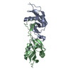
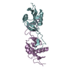
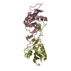
- Assembly
| Deposited unit | 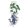
| ||||||||
|---|---|---|---|---|---|---|---|---|---|
| 1 |
| ||||||||
| Unit cell |
|
-Components
| #1: Protein | Mass: 15038.652 Da / Num. of mol.: 2 / Fragment: ligand-binding domain Source method: isolated from a genetically manipulated source Source: (gene. exp.) Homo sapiens (human) / Plasmid: pET28a / Species (production host): Escherichia coli / Production host:  Escherichia coli BL21(DE3) (bacteria) / Strain (production host): BL21(DE3) / References: UniProt: P78380 Escherichia coli BL21(DE3) (bacteria) / Strain (production host): BL21(DE3) / References: UniProt: P78380#2: Chemical | Ethylene glycol  Mass: 62.068 Da / Num. of mol.: 2 / Source method: obtained synthetically / Formula: C2H6O2 Mass: 62.068 Da / Num. of mol.: 2 / Source method: obtained synthetically / Formula: C2H6O2#3: Water | ChemComp-HOH / | Water Mass: 18.015 Da / Num. of mol.: 207 / Source method: isolated from a natural source / Formula: H2O Mass: 18.015 Da / Num. of mol.: 207 / Source method: isolated from a natural source / Formula: H2O |
|---|
-Experimental details
-Experiment
| Experiment | Method: X-RAY DIFFRACTION / Number of used crystals: 1 |
|---|
- Sample preparation
| Crystal | Density Matthews: 2.83 Å3/Da / Density % sol: 56.6 % |
|---|---|
| Crystal grow | Temperature: 293 K / Method: vapor diffusion, hanging drop / pH: 3.6 Details: Citrate, pH 3.6, VAPOR DIFFUSION, HANGING DROP, temperature 293K |
-Data collection
| Diffraction | Mean temperature: 100 K |
|---|---|
| Diffraction source | Source: SYNCHROTRON / Site: SPring-8  / Beamline: BL40B2 / Wavelength: 1 Å / Beamline: BL40B2 / Wavelength: 1 Å |
| Detector | Type: ADSC QUANTUM 4 / Detector: CCD / Date: Sep 27, 2003 |
| Radiation | Protocol: MAD / Monochromatic (M) / Laue (L): M / Scattering type: x-ray |
| Radiation wavelength | Wavelength: 1 Å / Relative weight: 1 |
| Reflection | Resolution: 1.78→50 Å / Num. obs: 33424 / % possible obs: 99.6 % / Biso Wilson estimate: 15.5 Å2 / Rmerge(I) obs: 0.053 |
- Processing
| Software |
| ||||||||||||||||||||||||||||||||||||
|---|---|---|---|---|---|---|---|---|---|---|---|---|---|---|---|---|---|---|---|---|---|---|---|---|---|---|---|---|---|---|---|---|---|---|---|---|---|
| Refinement | Method to determine structure: MAD / Resolution: 1.78→28.18 Å / Rfactor Rfree error: 0.006 / Data cutoff high absF: 279086.18 / Data cutoff low absF: 0 / Isotropic thermal model: RESTRAINED / Cross valid method: THROUGHOUT / σ(F): 0
| ||||||||||||||||||||||||||||||||||||
| Solvent computation | Solvent model: FLAT MODEL / Bsol: 47.5479 Å2 / ksol: 0.429383 e/Å3 | ||||||||||||||||||||||||||||||||||||
| Displacement parameters | Biso mean: 22.7 Å2
| ||||||||||||||||||||||||||||||||||||
| Refine analyze |
| ||||||||||||||||||||||||||||||||||||
| Refinement step | Cycle: LAST / Resolution: 1.78→28.18 Å
| ||||||||||||||||||||||||||||||||||||
| Refine LS restraints |
| ||||||||||||||||||||||||||||||||||||
| LS refinement shell | Resolution: 1.78→1.89 Å / Rfactor Rfree error: 0.018 / Total num. of bins used: 6
| ||||||||||||||||||||||||||||||||||||
| Xplor file |
|
