- PDB-1ywt: Crystal structure of the human sigma isoform of 14-3-3 in complex... -
+
データを開く
IDまたはキーワード:
読み込み中...
-
基本情報
登録情報
データベース: PDB / ID: 1ywt
タイトル
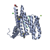
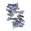
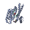
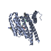
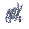
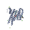
Crystal structure of the human sigma isoform of 14-3-3 in complex with a mode-1 phosphopeptide
要素
14-3-3 protein sigma
synthetic optimal phosphopeptide (mode-1)
キーワード
SIGNALING PROTEIN/DE NOVO PROTEIN / protein-phosphopeptide complex / 14-3-3 (14-3-3タンパク質) / SIGNALING PROTEIN-DE NOVO PROTEIN COMPLEX
機能・相同性
機能・相同性情報
regulation of epidermal cell division / protein kinase C inhibitor activity / positive regulation of epidermal cell differentiation / keratinocyte development / ケラチン / Regulation of localization of FOXO transcription factors / keratinocyte proliferation / phosphoserine residue binding / Activation of BAD and translocation to mitochondria / negative regulation of keratinocyte proliferation ...regulation of epidermal cell division / protein kinase C inhibitor activity / positive regulation of epidermal cell differentiation / keratinocyte development / ケラチン / Regulation of localization of FOXO transcription factors / keratinocyte proliferation / phosphoserine residue binding / Activation of BAD and translocation to mitochondria / negative regulation of keratinocyte proliferation / establishment of skin barrier / SARS-CoV-2 targets host intracellular signalling and regulatory pathways / Chk1/Chk2(Cds1) mediated inactivation of Cyclin B:Cdk1 complex / protein kinase A signaling / negative regulation of stem cell proliferation / SARS-CoV-1 targets host intracellular signalling and regulatory pathways / RHO GTPases activate PKNs / protein export from nucleus / negative regulation of innate immune response / protein sequestering activity / TP53 Regulates Transcription of Genes Involved in G2 Cell Cycle Arrest / release of cytochrome c from mitochondria / positive regulation of protein export from nucleus / stem cell proliferation / Translocation of SLC2A4 (GLUT4) to the plasma membrane / TP53 Regulates Metabolic Genes / negative regulation of protein kinase activity / negative regulation of cysteine-type endopeptidase activity involved in apoptotic process / intrinsic apoptotic signaling pathway in response to DNA damage / positive regulation of cell growth / regulation of cell cycle / cadherin binding / protein kinase binding / negative regulation of transcription by RNA polymerase II / シグナル伝達 / extracellular space / extracellular exosome / identical protein binding / 細胞核 / 細胞質基質 / 細胞質 類似検索 - 分子機能
SEQUENCE Chains C and D are an optimal phosphopeptide sequence as determined from a library screen, ...SEQUENCE Chains C and D are an optimal phosphopeptide sequence as determined from a library screen, and therefore have no database match.
 ムービー
ムービー コントローラー


 データを開く
 基本情報
基本情報 要素
要素 キーワード
キーワード SIGNALING PROTEIN/DE NOVO PROTEIN / protein-phosphopeptide complex /
  機能・相同性情報
 データ登録者
 引用
引用 構造の表示
構造の表示 ダウンロードとリンク
 その他のダウンロード


 PDBj
PDBj


 集合体
集合体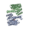


 分子量: 40.078 Da / 分子数: 1 / 由来タイプ: 合成 / 式: Ca
 分子量: 18.015 Da / 分子数: 55 / 由来タイプ: 天然 / 式: H2O
 試料調製
試料調製 / ビームライン: 8-BM / 波長: 0.97949 / 波長: 0.97949 Å
 解析
解析