 Movie
Movie Controller

[English] 日本語
 Yorodumi
- PDB-1yvk: Crystal Structure of the Bacillis subtilis Acetyltransferase in c... -
+ Open data

- Basic information
| Entry | Database: PDB / ID: 1yvk | ||||||
|---|---|---|---|---|---|---|---|
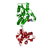
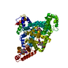
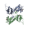
| Title | Crystal Structure of the Bacillis subtilis Acetyltransferase in complex with CoA, Northeast Structural Genomics Target SR237. | ||||||
 Components Components | hypothetical protein BSU33890 Hypothesis Hypothesis | ||||||
 Keywords Keywords | TRANSFERASE / alphs-beta protein / Structural Genomics / PSI / Protein Structure Initiative / Northeast Structural Genomics Consortium / NESG | ||||||
| Function / homology |  Function and homology informationN-acetyltransferase activity / Transferases; Acyltransferases; Transferring groups other than aminoacyl groups Function and homology informationN-acetyltransferase activity / Transferases; Acyltransferases; Transferring groups other than aminoacyl groupsSimilarity search - Function | ||||||
| Biological species |  Bacillus subtilis subsp. subtilis str. 168 (bacteria) Bacillus subtilis subsp. subtilis str. 168 (bacteria) | ||||||
| Method | X-RAY DIFFRACTION / SYNCHROTRON / SAD / Resolution: 3.01 Å | ||||||
 Authors Authors | Forouhar, F. / Yong, W. / Xiao, R. / Ciano, M. / Acton, T.B. / Montelione, G.T. / Hunt, J.F. / Tong, L. / Northeast Structural Genomics Consortium (NESG) | ||||||
 Citation Citation | Journal: To be Published Title: Crystal Structure of the Bacillis subtilis Acetyltransferase in complex with CoA, Northeast Structural Genomics Target SR237. Authors: Forouhar, F. / Yong, W. / Xiao, R. / Ciano, M. / Acton, T.B. / Montelione, G.T. / Hunt, J.F. / Tong, L. | ||||||
| History |
|
- Structure visualization
| Structure viewer | Molecule: MolmilJmol/JSmol |
|---|
- Downloads & links
-Download
| PDBx/mmCIF format | 1yvk.cif.gz | 130.2 KB | Display | PDBx/mmCIF format |
|---|---|---|---|---|
| PDB format | pdb1yvk.ent.gz | 110.1 KB | Display | PDB format |
| PDBx/mmJSON format | 1yvk.json.gz | Tree view | PDBx/mmJSON format | |
| Others |  Other downloads Other downloads |
-Validation report
| Arichive directory | https://data.pdbj.org/pub/pdb/validation_reports/yv/1yvkftp://data.pdbj.org/pub/pdb/validation_reports/yv/1yvk | HTTPS FTP |
|---|
-Related structure data
| Similar structure data | |
|---|---|
| Other databases |

-Links
 PDBj
PDBj


- Assembly
| Deposited unit | 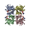
| ||||||||
|---|---|---|---|---|---|---|---|---|---|
| 1 |
| ||||||||
| Unit cell |
|
-Components
| #1: Protein | Hypothesis Mass: 18978.080 Da / Num. of mol.: 4 Source method: isolated from a genetically manipulated source Source: (gene. exp.) Bacillus subtilis subsp. subtilis str. 168 (bacteria)Species: Bacillus subtilis / Strain: subsp. subtilis str. 168 / Gene: yvbK / Plasmid: BL21 / Production host: Escherichia coli (E. coli) / Strain (production host): BL21(DE3)+MagicKeywords: Met substitution by Selno-Met and addition of C-tag (LEHHHHHH). YvbKReferences: UniProt: O32248 #2: Chemical | ChemComp-COA / Coenzyme A  Mass: 767.534 Da / Num. of mol.: 4 / Source method: obtained synthetically / Formula: C21H36N7O16P3S Mass: 767.534 Da / Num. of mol.: 4 / Source method: obtained synthetically / Formula: C21H36N7O16P3S |
|---|
-Experimental details
-Experiment
| Experiment | Method: X-RAY DIFFRACTION / Number of used crystals: 1 |
|---|
- Sample preparation
| Crystal | Density Matthews: 2.83 Å3/Da / Density % sol: 55 % |
|---|---|
| Crystal grow | Temperature: 293 K / Method: vapor diffusion, hanging drop / pH: 8.5 Details: 100 mM Tris-HCl (pH 8.5), 20% PEG 4K, 200 mM sodium acetate, 100 mM ammonium sulfate, 2 mM Acetyl-CoA, and 5 mM DTT., VAPOR DIFFUSION, HANGING DROP, temperature 293K |
-Data collection
| Diffraction | Mean temperature: 100 K |
|---|---|
| Diffraction source | Source: SYNCHROTRON / Site: NSLS  / Beamline: X4A / Wavelength: 0.97927 / Wavelength: 0.97927 Å / Beamline: X4A / Wavelength: 0.97927 / Wavelength: 0.97927 Å |
| Detector | Type: ADSC QUANTUM 4 / Detector: CCD / Date: Feb 7, 2005 / Details: mirrors |
| Radiation | Monochromator: Si 111 CHANNEL / Protocol: MAD / Monochromatic (M) / Laue (L): M / Scattering type: x-ray |
| Radiation wavelength | Wavelength: 0.97927 Å / Relative weight: 1 |
| Reflection | Resolution: 3→30 Å / Num. all: 29334 / Num. obs: 29334 / % possible obs: 95.7 % / Observed criterion σ(F): 0 / Observed criterion σ(I): 0 / Redundancy: 5.8 % / Biso Wilson estimate: 42.1 Å2 / Rmerge(I) obs: 0.108 / Rsym value: 0.075 / Net I/σ(I): 14.01 |
| Reflection shell | Resolution: 3→3.11 Å / Redundancy: 4.3 % / Rmerge(I) obs: 0.448 / Mean I/σ(I) obs: 2.32 / Num. unique all: 2388 / Rsym value: 0.343 / % possible all: 77.6 |
- Processing
| Software |
| |||||||||||||||||||||||||
|---|---|---|---|---|---|---|---|---|---|---|---|---|---|---|---|---|---|---|---|---|---|---|---|---|---|---|
| Refinement | Method to determine structure: SAD / Resolution: 3.01→30 Å / Rfactor Rfree error: 0.006 / Data cutoff high absF: 128262.73 / Data cutoff low absF: 0 / Isotropic thermal model: OVERALL / Cross valid method: THROUGHOUT / σ(F): 2 / σ(I): 2 / Stereochemistry target values: Engh & Huber
| |||||||||||||||||||||||||
| Solvent computation | Solvent model: FLAT MODEL / Bsol: 10 Å2 / ksol: 0.275366 e/Å3 | |||||||||||||||||||||||||
| Displacement parameters | Biso mean: 61.5 Å2
| |||||||||||||||||||||||||
| Refine analyze |
| |||||||||||||||||||||||||
| Refinement step | Cycle: LAST / Resolution: 3.01→30 Å
| |||||||||||||||||||||||||
| Refine LS restraints |
| |||||||||||||||||||||||||
| LS refinement shell | Resolution: 3.01→3.19 Å / Rfactor Rfree error: 0.03 / Total num. of bins used: 6
|
