 Movie
Movie Controller


+ Open data

- Basic information
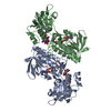
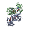
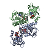
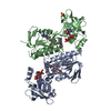
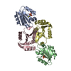
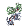
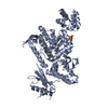
| Entry | Database: PDB / ID: 1yue | ||||||
|---|---|---|---|---|---|---|---|
| Title | Bacteriophage T4 capsid vertex protein gp24 | ||||||
 Components Components | Head vertex protein Gp24 | ||||||
 Keywords Keywords |  VIRAL PROTEIN / gp24 / bacteriophage T4 / capsid protein / vertex / bacteriophage / virus / HK97 / MAD VIRAL PROTEIN / gp24 / bacteriophage T4 / capsid protein / vertex / bacteriophage / virus / HK97 / MAD | ||||||
| Function / homology |  Function and homology information Function and homology information | ||||||
| Biological species |  Enterobacteria phage T4 (virus) Enterobacteria phage T4 (virus) | ||||||
| Method | X-RAY DIFFRACTION / SYNCHROTRON / MAD / Resolution: 2.9 Å | ||||||
 Authors Authors | Fokine, A. / Leiman, P.G. / Shneider, M.M. / Ahvazi, B. / Boeshans, K.M. / Steven, A.C. / Black, L.W. / Mesyanzhinov, V.V. / Rossmann, M.G. | ||||||
 Citation Citation | Journal: Proc.Natl.Acad.Sci.Usa / Year: 2005 Title: Structural and functional similarities between the capsid proteins of bacteriophages T4 and HK97 point to a common ancestry. Authors: Fokine, A. / Leiman, P.G. / Shneider, M.M. / Ahvazi, B. / Boeshans, K.M. / Steven, A.C. / Black, L.W. / Mesyanzhinov, V.V. / Rossmann, M.G. | ||||||
| History |
|
- Structure visualization
| Structure viewer | Molecule: MolmilJmol/JSmol |
|---|
- Downloads & links
-Download
| PDBx/mmCIF format | 1yue.cif.gz | 83.3 KB | Display | PDBx/mmCIF format |
|---|---|---|---|---|
| PDB format | pdb1yue.ent.gz | 67.5 KB | Display | PDB format |
| PDBx/mmJSON format | 1yue.json.gz | Tree view | PDBx/mmJSON format | |
| Others |  Other downloads Other downloads |
-Validation report
| Arichive directory | https://data.pdbj.org/pub/pdb/validation_reports/yu/1yueftp://data.pdbj.org/pub/pdb/validation_reports/yu/1yue | HTTPS FTP |
|---|
-Related structure data
| Similar structure data |
|---|

-Links
 PDBj
PDBj- Assembly
| Deposited unit | 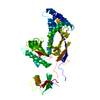
| ||||||||
|---|---|---|---|---|---|---|---|---|---|
| 1 |
| ||||||||
| Unit cell |
|
-Components
| #1: Protein | Mass: 47273.496 Da / Num. of mol.: 1 Source method: isolated from a genetically manipulated source Source: (gene. exp.) Enterobacteria phage T4 (virus) / Genus: T4-like viruses / Species: Enterobacteria phage T4 sensu lato / Gene: 24 / Plasmid: pET-23 / Production host:  Escherichia coli (E. coli) / Strain (production host): B834(DE3) / References: UniProt: P19896 Escherichia coli (E. coli) / Strain (production host): B834(DE3) / References: UniProt: P19896 |
|---|
-Experimental details
-Experiment
| Experiment | Method: X-RAY DIFFRACTION / Number of used crystals: 1 |
|---|
- Sample preparation
| Crystal | Density Matthews: 3.7 Å3/Da / Density % sol: 0.63 % |
|---|---|
| Crystal grow | Temperature: 293 K / Method: vapor diffusion, sitting drop / pH: 7.5 Details: 0.1 M HEPES-Na pH 7.5; 10% v/v isopropanol; 0.2 M Sodium Citrate, VAPOR DIFFUSION, SITTING DROP, temperature 293K |
-Data collection
| Diffraction |
| ||||||||||||||||||
|---|---|---|---|---|---|---|---|---|---|---|---|---|---|---|---|---|---|---|---|
| Diffraction source |
| ||||||||||||||||||
| Detector |
| ||||||||||||||||||
| Radiation | Protocol: MAD / Monochromatic (M) / Laue (L): M / Scattering type: x-ray | ||||||||||||||||||
| Radiation wavelength |
| ||||||||||||||||||
| Reflection | Resolution: 2.9→35 Å / Num. all: 164088 / Num. obs: 11621 / % possible obs: 97.7 % / Observed criterion σ(F): 2 / Observed criterion σ(I): 2 / Redundancy: 14.2 % / Biso Wilson estimate: 108 Å2 / Rmerge(I) obs: 0.073 / Net I/σ(I): 24.6 |

- Processing
| Software |
| |||||||||||||||||||||||||
|---|---|---|---|---|---|---|---|---|---|---|---|---|---|---|---|---|---|---|---|---|---|---|---|---|---|---|
| Refinement | Method to determine structure: MAD / Resolution: 2.9→35 Å / Isotropic thermal model: isotropic / Cross valid method: THROUGHOUT / σ(F): 2 / Stereochemistry target values: Engh & Huber Details: High values of atomic B factors reflect the disorder of the crystal. The mosaisity was ~1.5 deg
| |||||||||||||||||||||||||
| Solvent computation | Solvent model: Flat Model / Bsol: 48.1183 Å2 / ksol: 0.30571 e/Å3 | |||||||||||||||||||||||||
| Displacement parameters | Biso mean: 108.7 Å2
| |||||||||||||||||||||||||
| Refine analyze |
| |||||||||||||||||||||||||
| Refinement step | Cycle: LAST / Resolution: 2.9→35 Å
| |||||||||||||||||||||||||
| Refine LS restraints |
|
