 Movie
Movie Controller


+ Open data

- Basic information
| Entry | Database: PDB / ID: 1ypy | ||||||
|---|---|---|---|---|---|---|---|
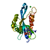
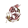
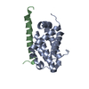
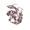
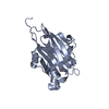
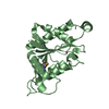
| Title | Crystal Structure of Vaccinia Virus L1 protein | ||||||
 Components Components | Virion membrane protein | ||||||
 Keywords Keywords |  VIRAL PROTEIN / Vaccinia virus / Orthopox / Variola virus / smallpox / L1 VIRAL PROTEIN / Vaccinia virus / Orthopox / Variola virus / smallpox / L1 | ||||||
| Function / homology | Virion membrane protein, poxvirus L1-related / Lipid membrane protein of large eukaryotic DNA viruses / symbiont entry into host cell / viral envelope / virion attachment to host cell / virion membrane / membrane / Entry-fusion complex associated protein OPG095 Function and homology information Function and homology information | ||||||
| Biological species |  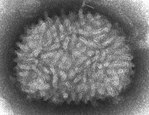 Vaccinia virus Vaccinia virus | ||||||
| Method | X-RAY DIFFRACTION / SYNCHROTRON / SAD / Resolution: 1.51 Å | ||||||
 Authors Authors | Su, H.P. / Garman, S.C. / Allison, T.J. / Fogg, C. / Moss, B. / Garboczi, D.N. | ||||||
 Citation Citation | Journal: Proc.Natl.Acad.Sci.Usa / Year: 2005 Title: The 1.51-Angstrom structure of the poxvirus L1 protein, a target of potent neutralizing antibodies. Authors: Su, H.P. / Garman, S.C. / Allison, T.J. / Fogg, C. / Moss, B. / Garboczi, D.N. | ||||||
| History |
|
- Structure visualization
| Structure viewer | Molecule: MolmilJmol/JSmol |
|---|
- Downloads & links
-Download
| PDBx/mmCIF format | 1ypy.cif.gz | 80.6 KB | Display | PDBx/mmCIF format |
|---|---|---|---|---|
| PDB format | pdb1ypy.ent.gz | 64 KB | Display | PDB format |
| PDBx/mmJSON format | 1ypy.json.gz | Tree view | PDBx/mmJSON format | |
| Others |  Other downloads Other downloads |
-Validation report
| Arichive directory | https://data.pdbj.org/pub/pdb/validation_reports/yp/1ypyftp://data.pdbj.org/pub/pdb/validation_reports/yp/1ypy | HTTPS FTP |
|---|
-Related structure data
| Similar structure data |
|---|


-Links
 PDBj
PDBj- Assembly
| Deposited unit | 
| ||||||||
|---|---|---|---|---|---|---|---|---|---|
| 1 | 
| ||||||||
| 2 | 
| ||||||||
| Unit cell |
|
-Components
| #1: Protein | Mass: 19532.877 Da / Num. of mol.: 2 Source method: isolated from a genetically manipulated source Source: (gene. exp.) Vaccinia virus / Genus: Orthopoxvirus / Gene: L1 / Plasmid: pLM / Production host:  Escherichia coli (E. coli) / Strain (production host): BL21(DE3)-CodonPlus-RIL / References: UniProt: P07612 Escherichia coli (E. coli) / Strain (production host): BL21(DE3)-CodonPlus-RIL / References: UniProt: P07612#2: Water | ChemComp-HOH / | Water Mass: 18.015 Da / Num. of mol.: 327 / Source method: isolated from a natural source / Formula: H2O Mass: 18.015 Da / Num. of mol.: 327 / Source method: isolated from a natural source / Formula: H2O |
|---|
-Experimental details
-Experiment
| Experiment | Method: X-RAY DIFFRACTION / Number of used crystals: 2 |
|---|
- Sample preparation
| Crystal |
| |||||||||||||||
|---|---|---|---|---|---|---|---|---|---|---|---|---|---|---|---|---|
| Crystal grow |
|
-Data collection
| Diffraction |
| |||||||||||||||
|---|---|---|---|---|---|---|---|---|---|---|---|---|---|---|---|---|
| Diffraction source |
| |||||||||||||||
| Detector |
| |||||||||||||||
| Radiation |
| |||||||||||||||
| Radiation wavelength |
| |||||||||||||||
| Reflection | Number: 15507 | |||||||||||||||
| Reflection | Resolution: 1.51→50 Å / Num. obs: 52912 / % possible obs: 99.1 % / Observed criterion σ(F): 0 / Observed criterion σ(I): 0 / Redundancy: 3.7 % / Biso Wilson estimate: 23.5 Å2 / Rmerge(I) obs: 0.064 / Χ2: 1.058 / Net I/σ(I): 12.3 | |||||||||||||||
| Reflection shell | Resolution: 1.51→1.56 Å / Redundancy: 3.7 % / Rmerge(I) obs: 0.496 / Mean I/σ(I) obs: 3.55 / Num. unique all: 5290 / Χ2: 1.045 / % possible all: 100 |

-Phasing
| Phasing | Method: SAD | |||||||||||||||||||||||||||||||||||||||||||||||||
|---|---|---|---|---|---|---|---|---|---|---|---|---|---|---|---|---|---|---|---|---|---|---|---|---|---|---|---|---|---|---|---|---|---|---|---|---|---|---|---|---|---|---|---|---|---|---|---|---|---|---|
| Phasing MAD set site |
| |||||||||||||||||||||||||||||||||||||||||||||||||
| Phasing dm | FOM : 0.74 / FOM acentric: 0.76 / FOM centric: 0.57 / Reflection: 6895 / Reflection acentric: 6398 / Reflection centric: 497 | |||||||||||||||||||||||||||||||||||||||||||||||||
| Phasing dm shell |
|
- Processing
| Software |
| ||||||||||||||||||||||||||||
|---|---|---|---|---|---|---|---|---|---|---|---|---|---|---|---|---|---|---|---|---|---|---|---|---|---|---|---|---|---|
| Refinement | Method to determine structure: SAD / Resolution: 1.51→50 Å / Data cutoff high absF: 10000 / Data cutoff low absF: 0 / σ(F): 0 / Stereochemistry target values: Engh & Huber Details: The close contact between B5 (CB ALA) and B82 (CG GLU) may indicate flexibility in side chain of B82 as well as the disorder in B5, which is the n-terminus in that molecule. It is possible ...Details: The close contact between B5 (CB ALA) and B82 (CG GLU) may indicate flexibility in side chain of B82 as well as the disorder in B5, which is the n-terminus in that molecule. It is possible that there are alternate positions in both residues to support packing between the asymmetric units at this interface, but the model is the best fit that did not violate geometric requirements.
| ||||||||||||||||||||||||||||
| Displacement parameters | Biso mean: 58.345 Å2
| ||||||||||||||||||||||||||||
| Refine analyze |
| ||||||||||||||||||||||||||||
| Refinement step | Cycle: LAST / Resolution: 1.51→50 Å
| ||||||||||||||||||||||||||||
| Refine LS restraints |
| ||||||||||||||||||||||||||||
| LS refinement shell | Resolution: 1.51→1.6 Å / Rfactor Rfree error: 0.015
|
