 Movie
Movie Controller

[English] 日本語
 Yorodumi
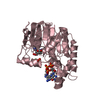
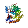
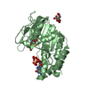
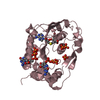
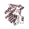
- PDB-1yp0: Structure of the steroidogenic factor-1 ligand binding domain bou... -
+ Open data

- Basic information
| Entry | Database: PDB / ID: 1yp0 | ||||||
|---|---|---|---|---|---|---|---|
| Title | Structure of the steroidogenic factor-1 ligand binding domain bound to phospholipid and a SHP peptide motif | ||||||
 Components Components |
| ||||||
 Keywords Keywords |  TRANSCRIPTION / Ligand dependent nuclear receptor / Steroidogenic factor-1 / NR5A1 / helical sandwich fold / LBD fold TRANSCRIPTION / Ligand dependent nuclear receptor / Steroidogenic factor-1 / NR5A1 / helical sandwich fold / LBD fold | ||||||
| Function / homology |  Function and homology information Function and homology informationresponse to gonadotropin-releasing hormone / Sertoli cell differentiation / reproductive process / SUMOylation of intracellular receptors / negative regulation of female gonad development / Nuclear Receptor transcription pathway / sex determination / positive regulation of male gonad development / luteinization / calcineurin-mediated signaling ...response to gonadotropin-releasing hormone / Sertoli cell differentiation / reproductive process / SUMOylation of intracellular receptors / negative regulation of female gonad development / Nuclear Receptor transcription pathway / sex determination / positive regulation of male gonad development / luteinization / calcineurin-mediated signaling / Nuclear Receptor transcription pathway / tissue development / Leydig cell differentiation / male sex determination / nuclear retinoic acid receptor binding / maintenance of protein location in nucleus / bile acid and bile salt transport / hormone metabolic process / transcription regulator inhibitor activity / peroxisome proliferator activated receptor binding / nuclear thyroid hormone receptor binding / adrenal gland development / female gonad development / animal organ regeneration / response to glucose / nuclear retinoid X receptor binding / Notch signaling pathway / hormone-mediated signaling pathway / transcription coregulator binding / RNA polymerase II transcription regulatory region sequence-specific DNA binding / phospholipid binding / circadian regulation of gene expression / positive regulation of insulin secretion / negative regulation of DNA-binding transcription factor activity / response to organic cyclic compound / circadian rhythm / RNA polymerase II transcription regulator complex / male gonad development / transcription corepressor activity / nuclear receptor activity / sequence-specific double-stranded DNA binding / double-stranded DNA binding / response to ethanol / sequence-specific DNA binding / transcription by RNA polymerase II / DNA-binding transcription factor activity, RNA polymerase II-specific / RNA polymerase II cis-regulatory region sequence-specific DNA binding / DNA-binding transcription factor activity / protein domain specific binding / negative regulation of gene expression / negative regulation of DNA-templated transcription / chromatin binding / chromatin / protein-containing complex binding / positive regulation of gene expression / regulation of DNA-templated transcription / regulation of transcription by RNA polymerase II / positive regulation of DNA-templated transcription / negative regulation of transcription by RNA polymerase II / enzyme binding / protein homodimerization activity / positive regulation of transcription by RNA polymerase II / protein-containing complex / DNA binding / zinc ion binding / nucleoplasm / nucleus / cytosol / cytoplasmSimilarity search - Function | ||||||
| Biological species |  Mus musculus (house mouse) Mus musculus (house mouse) | ||||||
| Method | X-RAY DIFFRACTION / SYNCHROTRON / MOLECULAR REPLACEMENT / Resolution: 1.5 Å | ||||||
 Authors Authors | Li, Y. / Choi, M. / Cavey, G. / Daugherty, J. / Suino, K. / Kovach, A. / Bingham, N. / Kliewer, S. / Xu, H. | ||||||
 Citation Citation | Journal: Mol.Cell / Year: 2005 Title: Crystallographic identification and functional characterization of phospholipids as ligands for the orphan nuclear receptor steroidogenic factor-1. Authors: Li, Y. / Choi, M. / Cavey, G. / Daugherty, J. / Suino, K. / Kovach, A. / Bingham, N. / Kliewer, S. / Xu, H. | ||||||
| History |
|
- Structure visualization
| Structure viewer | Molecule: MolmilJmol/JSmol |
|---|
- Downloads & links
-Download
| PDBx/mmCIF format | 1yp0.cif.gz | 68.7 KB | Display | PDBx/mmCIF format |
|---|---|---|---|---|
| PDB format | pdb1yp0.ent.gz | 50.3 KB | Display | PDB format |
| PDBx/mmJSON format | 1yp0.json.gz | Tree view | PDBx/mmJSON format | |
| Others |  Other downloads Other downloads |
-Validation report
| Arichive directory | https://data.pdbj.org/pub/pdb/validation_reports/yp/1yp0ftp://data.pdbj.org/pub/pdb/validation_reports/yp/1yp0 | HTTPS FTP |
|---|
-Related structure data
| Similar structure data |
|---|


-Links
 PDBj
PDBj


- Assembly
| Deposited unit | 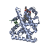
| ||||||||
|---|---|---|---|---|---|---|---|---|---|
| 1 |
| ||||||||
| Unit cell |
|
-Components
| #1: Protein | Mass: 27513.010 Da / Num. of mol.: 1 / Fragment: ligand binding domain (Residues: 223-461) Source method: isolated from a genetically manipulated source Source: (gene. exp.) Mus musculus (house mouse) / Plasmid: PET24 / Species (production host): Escherichia coli / Production host:  Escherichia coli BL21(DE3) (bacteria) / Strain (production host): BL21-DE3 / References: UniProt: P33242 Escherichia coli BL21(DE3) (bacteria) / Strain (production host): BL21-DE3 / References: UniProt: P33242 |
|---|---|
| #2: Protein/peptide | Mass: 1312.534 Da / Num. of mol.: 1 / Fragment: SHP LXXLL / Source method: obtained synthetically Details: peptide synthesis, the sequence of the peptide is naturally found in Rattus norvegicus (Norway rat) References: UniProt: P97947 |
| #3: Chemical | ChemComp-PEF / Phosphatidylethanolamine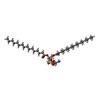  Mass: 691.959 Da / Num. of mol.: 1 / Source method: obtained synthetically / Formula: C37H74NO8P / Comment: phospholipid*YM Mass: 691.959 Da / Num. of mol.: 1 / Source method: obtained synthetically / Formula: C37H74NO8P / Comment: phospholipid*YM |
| #4: Water | ChemComp-HOH / Water Mass: 18.015 Da / Num. of mol.: 196 / Source method: isolated from a natural source / Formula: H2O Mass: 18.015 Da / Num. of mol.: 196 / Source method: isolated from a natural source / Formula: H2O |
-Experimental details
-Experiment
| Experiment | Method: X-RAY DIFFRACTION / Number of used crystals: 1 |
|---|
- Sample preparation
| Crystal | Density Matthews: 2.6 Å3/Da / Density % sol: 52 % |
|---|---|
| Crystal grow | Temperature: 293 K / Method: vapor diffusion, hanging drop / pH: 6 Details: PEG3350, pH 6.0, VAPOR DIFFUSION, HANGING DROP, temperature 293K |
-Data collection
| Diffraction | Mean temperature: 100 K |
|---|---|
| Diffraction source | Source: SYNCHROTRON / Site: APS  / Beamline: 32-ID / Wavelength: 1 Å / Beamline: 32-ID / Wavelength: 1 Å |
| Detector | Type: MARRESEARCH / Detector: CCD / Date: Oct 21, 2004 |
| Radiation | Monochromator: diamond crystal / Protocol: SINGLE WAVELENGTH / Monochromatic (M) / Laue (L): M / Scattering type: x-ray |
| Radiation wavelength | Wavelength: 1 Å / Relative weight: 1 |
| Reflection | Resolution: 1.5→30 Å / Num. all: 51068 / Num. obs: 51068 / % possible obs: 100 % / Observed criterion σ(F): 0 / Observed criterion σ(I): 0 / Redundancy: 10 % / Biso Wilson estimate: 22.9 Å2 / Rmerge(I) obs: 0.125 / Rsym value: 0.125 / Net I/σ(I): 34.8 |
| Reflection shell | Resolution: 1.5→1.55 Å / Redundancy: 6 % / Mean I/σ(I) obs: 3 / % possible all: 100 |
- Processing
| Software |
| ||||||||||||||||||||||||||||||||||||||||||||||||||||||||||||
|---|---|---|---|---|---|---|---|---|---|---|---|---|---|---|---|---|---|---|---|---|---|---|---|---|---|---|---|---|---|---|---|---|---|---|---|---|---|---|---|---|---|---|---|---|---|---|---|---|---|---|---|---|---|---|---|---|---|---|---|---|---|
| Refinement | Method to determine structure: MOLECULAR REPLACEMENT Starting model: mLRH-1/SHP complex Resolution: 1.5→10 Å / Rfactor Rfree error: 0.004 / Data cutoff high absF: 1729788.93 / Data cutoff low absF: 0 / Isotropic thermal model: RESTRAINED / Cross valid method: THROUGHOUT / σ(F): 0 / Stereochemistry target values: Engh & Huber
| ||||||||||||||||||||||||||||||||||||||||||||||||||||||||||||
| Solvent computation | Solvent model: FLAT MODEL / Bsol: 67.1241 Å2 / ksol: 0.498201 e/Å3 | ||||||||||||||||||||||||||||||||||||||||||||||||||||||||||||
| Displacement parameters | Biso mean: 29.1 Å2
| ||||||||||||||||||||||||||||||||||||||||||||||||||||||||||||
| Refine analyze |
| ||||||||||||||||||||||||||||||||||||||||||||||||||||||||||||
| Refinement step | Cycle: LAST / Resolution: 1.5→10 Å
| ||||||||||||||||||||||||||||||||||||||||||||||||||||||||||||
| Refine LS restraints |
| ||||||||||||||||||||||||||||||||||||||||||||||||||||||||||||
| LS refinement shell | Resolution: 1.5→1.59 Å / Rfactor Rfree error: 0.011 / Total num. of bins used: 6
| ||||||||||||||||||||||||||||||||||||||||||||||||||||||||||||
| Xplor file |
|
