[English] 日本語
Yorodumi
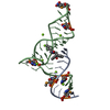
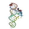
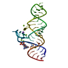
- PDB-1ykv: Crystal structure of the Diels-Alder ribozyme complexed with the ... -

+
Open data


ID or keywords:

Loading...

-
Basic information

Entry
Database: PDB / ID: 1ykv
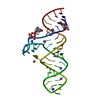
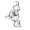
TitleCrystal structure of the Diels-Alder ribozyme complexed with the product of the reaction between N-pentylmaleimide and covalently attached 9-hydroxymethylanthracene
Components(Diels-Alder ribozyme) x 2
KeywordsRNA / carbon-carbon bond / catalytic mechanism / Diels-Alder reaction / ribozyme
Function / homologyChem-DAI / RNA / RNA (> 10)
Function and homology information
MethodX-RAY DIFFRACTION / SYNCHROTRON / MOLECULAR REPLACEMENT / Resolution: 3.3 Å
AuthorsSerganov, A. / Keiper, S. / Malinina, L. / Tereshko, V. / Skripkin, E. / Hobartner, C. / Polonskaia, A. / Phan, A.T. / Wombacher, R. / Micura, R. ...Serganov, A. / Keiper, S. / Malinina, L. / Tereshko, V. / Skripkin, E. / Hobartner, C. / Polonskaia, A. / Phan, A.T. / Wombacher, R. / Micura, R. / Dauter, Z. / Jaschke, A. / Patel, D.J.
CitationJournal: Nat.Struct.Mol.Biol. / Year: 2005
Title: Structural basis for Diels-Alder ribozyme-catalyzed carbon-carbon bond formation.
Authors: Serganov, A. / Keiper, S. / Malinina, L. / Tereshko, V. / Skripkin, E. / Hobartner, C. / Polonskaia, A. / Phan, A.T. / Wombacher, R. / Micura, R. / Dauter, Z. / Jaschke, A. / Patel, D.J.
History
DepositionJan 18, 2005Deposition site: RCSB / Processing site: RCSB
Revision 1.0Feb 22, 2005Provider: repository / Type: Initial release
Revision 1.1Apr 30, 2008Group: Version format compliance
Revision 1.2Jul 13, 2011Group: Advisory / Refinement description / Version format compliance
Revision 1.3Aug 23, 2023Group: Data collection / Database references ...Data collection / Database references / Derived calculations / Refinement description
Category: chem_comp_atom / chem_comp_bond ...chem_comp_atom / chem_comp_bond / database_2 / pdbx_initial_refinement_model / struct_conn / struct_ncs_dom_lim / struct_site
Item: _database_2.pdbx_DOI / _database_2.pdbx_database_accession ..._database_2.pdbx_DOI / _database_2.pdbx_database_accession / _struct_conn.pdbx_dist_value / _struct_conn.ptnr1_auth_comp_id / _struct_conn.ptnr1_auth_seq_id / _struct_conn.ptnr1_label_asym_id / _struct_conn.ptnr1_label_atom_id / _struct_conn.ptnr1_label_comp_id / _struct_conn.ptnr1_label_seq_id / _struct_conn.ptnr2_auth_comp_id / _struct_conn.ptnr2_auth_seq_id / _struct_conn.ptnr2_label_asym_id / _struct_conn.ptnr2_label_atom_id / _struct_conn.ptnr2_label_comp_id / _struct_conn.ptnr2_label_seq_id / _struct_ncs_dom_lim.beg_auth_comp_id / _struct_ncs_dom_lim.beg_label_asym_id / _struct_ncs_dom_lim.beg_label_seq_id / _struct_ncs_dom_lim.end_auth_comp_id / _struct_site.pdbx_auth_asym_id / _struct_site.pdbx_auth_comp_id / _struct_site.pdbx_auth_seq_id
Remark 999SEQUENCE The DAI hetgroup is connected to the 5' G of the 11-mer RNA via hexaethyleneglycol linker. ...SEQUENCE The DAI hetgroup is connected to the 5' G of the 11-mer RNA via hexaethyleneglycol linker. The linker is not visible in the electron density.

-
Structure visualization

Structure viewerMolecule:
MolmilJmol/JSmol

Downloads & links

-
Assembly

Deposited unit
A: Diels-Alder ribozyme
B: Diels-Alder ribozyme
C: Diels-Alder ribozyme
D: Diels-Alder ribozyme
hetero molecules


Theoretical massNumber of molelcules
Total (without water)32,44013
Polymers31,5194
Non-polymers9219
Water0
1
A: Diels-Alder ribozyme
B: Diels-Alder ribozyme
hetero molecules


Theoretical massNumber of molelcules
Total (without water)16,2327
Polymers15,7592
Non-polymers4735
Water0
TypeNameSymmetry operationNumber
identity operation1_555x,y,z1
2
C: Diels-Alder ribozyme
D: Diels-Alder ribozyme
hetero molecules


Theoretical massNumber of molelcules
Total (without water)16,2086
Polymers15,7592
Non-polymers4484
Water0
TypeNameSymmetry operationNumber
identity operation1_555x,y,z1
Unit cell
Length a, b, c (Å)78.288, 44.039, 79.998
Angle α, β, γ (deg.)90.00, 106.87, 90.00
Int Tables number4
Space group name H-MP1211
Noncrystallographic symmetry (NCS)NCS domain:
IDEns-IDDetails
11A
21C
12B
22D

NCS domain segments:

Component-ID: 1 / End auth comp-ID: C / End label comp-ID: C / Refine code: 6

Dom-IDEns-IDBeg auth comp-IDBeg label comp-IDEnd label seq-IDAuth asym-IDLabel asym-IDAuth seq-IDLabel seq-ID
11DAIDAI11AE - A100 - 111
21DAIDAI11CJ - C100 - 111
12GGBB201 - 2381 - 38
22GGDD201 - 2381 - 38

NCS ensembles :
ID
1
2

-
Components

#1: RNA chain Diels-Alder ribozyme


Mass: 3497.146 Da / Num. of mol.: 2 / Source method: obtained synthetically
Details: This RNA was chemically synthesized. The sequence was derived from the in vitro selected ribozyme catalysing Diels-Alder reaction between thethered anthracene and biotinylated maleimide
#2: RNA chain Diels-Alder ribozyme


Mass: 12262.333 Da / Num. of mol.: 2 / Source method: obtained synthetically
Details: This RNA was chemically synthesized. The sequence was derived from the in vitro selected ribozyme catalysing Diels-Alder reaction between thethered anthracene and biotinylated maleimide
#3: Chemical ChemComp-DAI / (3AS,9AS)-2-PENTYL-4-HYDROXYMETHYL-3A,4,9,9A-TETRAHYDRO-4,9[1',2']-BENZENO-1H-BENZ[F]ISOINDOLE-1,3(2H)-DIONE / (11S,12S)-9-HYDROXYMETHYL-11,12-(2-PENTYL-1,3-DIOXO-2-AZAPROPANO)-9,10-DIHYDRO-9,10-ETHANOANTHRACENE


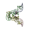
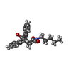
Mass: 375.460 Da / Num. of mol.: 2 / Source method: obtained synthetically / Formula: C24H25NO3
#4: Chemical
ChemComp-MG / MAGNESIUM ION


Mass: 24.305 Da / Num. of mol.: 7 / Source method: obtained synthetically / Formula: Mg

-
Experimental details

-
Experiment

ExperimentMethod: X-RAY DIFFRACTION / Number of used crystals: 1

-
Sample preparation

CrystalDensity Matthews: 3.99 Å3/Da / Density % sol: 63 %
Crystal growTemperature: 293 K / Method: vapor diffusion, hanging drop / pH: 7.5
Details: PEG400, sodium HEPES, magnesium chloride, ethylacetate, pH 7.5, VAPOR DIFFUSION, HANGING DROP, temperature 293K
Components of the solutions
IDNameCrystal-IDSol-ID
1PEG40011
2sodium HEPES11
3H2O11
4PEG40012
5sodium HEPES12
6magnesium chloride12
7ethylacetate12
8H2O12

-
Data collection

DiffractionMean temperature: 100 K
Diffraction sourceSource: SYNCHROTRON / Site: APS / Beamline: 14-BM-D / Wavelength: 1.1 Å
DetectorType: ADSC QUANTUM 4 / Detector: CCD / Date: Oct 3, 2002
Details: Si (111) double-crystal monochromator, Bent cylindrical Si-mirror (Rh coating)
RadiationMonochromator: Si (111) double-crystal monochromator / Protocol: SINGLE WAVELENGTH / Monochromatic (M) / Laue (L): M / Scattering type: x-ray
Radiation wavelengthWavelength: 1.1 Å / Relative weight: 1
ReflectionResolution: 3.3→20 Å / Num. all: 8607 / Num. obs: 8392 / % possible obs: 97.7 % / Observed criterion σ(F): 0 / Observed criterion σ(I): 0 / Redundancy: 4.9 % / Rmerge(I) obs: 0.092 / Net I/σ(I): 14
Reflection shellResolution: 3.3→3.42 Å / Redundancy: 4.4 % / Rmerge(I) obs: 0.162 / Mean I/σ(I) obs: 8 / % possible all: 88.9

-
Processing

Software
NameVersionClassification
REFMAC5.1.9999refinement
HKL-2000data reduction
SCALEPACKdata scaling
AMoREphasing
RefinementMethod to determine structure: MOLECULAR REPLACEMENT
Starting model: The Diels-Alder ribozyme structure from the Diels-Alder ribozyme-product complex, pdb entry 1YLS
Resolution: 3.3→20 Å / Cor.coef. Fo:Fc: 0.899 / Cor.coef. Fo:Fc free: 0.862 / SU B: 53.158 / SU ML: 0.383 / TLS residual ADP flag: LIKELY RESIDUAL / Cross valid method: THROUGHOUT / σ(F): 0 / σ(I): 0 / ESU R Free: 0.474 / Stereochemistry target values: MAXIMUM LIKELIHOOD / Details: HYDROGENS HAVE BEEN ADDED IN THE RIDING POSITIONS
RfactorNum. reflection% reflectionSelection details
Rfree0.26206 388 4.9 %RANDOM
Rwork0.22802 ---
all0.22971 7910 --
obs0.22971 7522 97.7 %-
Solvent computationIon probe radii: 0.8 Å / Shrinkage radii: 0.8 Å / VDW probe radii: 1.2 Å / Solvent model: MASK
Displacement parametersBiso mean: 21.884 Å2
Baniso -1Baniso -2Baniso -3
1-3.52 Å20 Å2-0.82 Å2
2---3.78 Å20 Å2
3----0.22 Å2
Refine analyzeLuzzati coordinate error obs: 0.607 Å
Refinement stepCycle: LAST / Resolution: 3.3→20 Å
ProteinNucleic acidLigandSolventTotal
Num. atoms0 2092 63 0 2155
Refine LS restraints
Refine-IDTypeDev idealDev ideal targetNumber
X-RAY DIFFRACTIONr_bond_refined_d0.0160.0212404
X-RAY DIFFRACTIONr_bond_other_d0.0060.02846
X-RAY DIFFRACTIONr_angle_refined_deg1.4833734
X-RAY DIFFRACTIONr_angle_other_deg2.59732200
X-RAY DIFFRACTIONr_chiral_restr0.0940.2496
X-RAY DIFFRACTIONr_gen_planes_refined0.0090.021074
X-RAY DIFFRACTIONr_gen_planes_other0.0090.0216
X-RAY DIFFRACTIONr_nbd_refined0.1680.2593
X-RAY DIFFRACTIONr_nbd_other0.310.21154
X-RAY DIFFRACTIONr_nbtor_other0.1150.2589
X-RAY DIFFRACTIONr_xyhbond_nbd_refined0.1410.246
X-RAY DIFFRACTIONr_xyhbond_nbd_other0.3690.22
X-RAY DIFFRACTIONr_symmetry_vdw_refined0.2060.240
X-RAY DIFFRACTIONr_symmetry_vdw_other0.2230.211
X-RAY DIFFRACTIONr_symmetry_hbond_refined0.130.23
X-RAY DIFFRACTIONr_scbond_it0.63933432
X-RAY DIFFRACTIONr_scangle_it0.5994.53734
Refine LS restraints NCS

Dom-ID: 1 / Refine-ID: X-RAY DIFFRACTION

Ens-IDAuth asym-IDNumberTypeRms dev position (Å)Weight position
1A376loose positional0.35
2B1122loose positional0.385
1A376loose thermal0.410
2B1122loose thermal0.4410
LS refinement shellResolution: 3.3→3.383 Å / Total num. of bins used: 20 /
RfactorNum. reflection
Rfree0.297 35
Rwork0.326 568
Refinement TLS params.

Method: refined / Refine-ID: X-RAY DIFFRACTION

IDL112)L122)L132)L222)L232)L332)S11 (Å °)S12 (Å °)S13 (Å °)S21 (Å °)S22 (Å °)S23 (Å °)S31 (Å °)S32 (Å °)S33 (Å °)T112)T122)T132)T222)T232)T332)Origin x (Å)Origin y (Å)Origin z (Å)
16.6386-0.1901-7.88452.694-1.182410.1018-0.03290.45670.38020.12150.61920.46550.032-0.8974-0.5863-0.0677-0.01680.2104-0.19840.1709-0.196228.294225.601234.0195
21.0523-0.402-1.31014.03710.68615.9779-0.03550.1519-0.0768-0.43170.28390.34780.2254-0.0508-0.2484-0.2742-0.1219-0.0152-0.30250.0803-0.337537.821826.750919.6474
310.113-0.68078.47522.29981.84779.6968-0.17070.48620.21520.10660.4069-0.53090.12340.7441-0.2362-0.0752-0.0343-0.2325-0.2747-0.15670.088365.8634.28133.9042
41.213-1.63981.91513.108-1.08095.57590.03320.39280.1433-0.38020.231-0.5544-0.09750.4293-0.2642-0.2679-0.1455-0.0531-0.1949-0.0477-0.119856.240133.404319.605
Refinement TLS group
IDRefine-IDRefine TLS-IDAuth asym-IDAuth seq-ID
1X-RAY DIFFRACTION1A101 - 111
2X-RAY DIFFRACTION2B201 - 238
3X-RAY DIFFRACTION3C101 - 111
4X-RAY DIFFRACTION4D201 - 238

+
About Yorodumi

-
News

-
Feb 9, 2022. New format data for meta-information of EMDB entries

New format data for meta-information of EMDB entries

  • Version 3 of the EMDB header file is now the official format.
  • The previous official version 1.9 will be removed from the archive.

Related info.:EMDB header

External links:wwPDB to switch to version 3 of the EMDB data model

-
Aug 12, 2020. Covid-19 info

Covid-19 info

URL: https://pdbj.org/emnavi/covid19.php

New page: Covid-19 featured information page in EM Navigator.

Related info.:Covid-19 info / Mar 5, 2020. Novel coronavirus structure data

+
Mar 5, 2020. Novel coronavirus structure data

Novel coronavirus structure data

Related info.:Yorodumi Speices / Aug 12, 2020. Covid-19 info

External links:COVID-19 featured content - PDBj / Molecule of the Month (242):Coronavirus Proteases

+
Jan 31, 2019. EMDB accession codes are about to change! (news from PDBe EMDB page)

EMDB accession codes are about to change! (news from PDBe EMDB page)

  • The allocation of 4 digits for EMDB accession codes will soon come to an end. Whilst these codes will remain in use, new EMDB accession codes will include an additional digit and will expand incrementally as the available range of codes is exhausted. The current 4-digit format prefixed with “EMD-” (i.e. EMD-XXXX) will advance to a 5-digit format (i.e. EMD-XXXXX), and so on. It is currently estimated that the 4-digit codes will be depleted around Spring 2019, at which point the 5-digit format will come into force.
  • The EM Navigator/Yorodumi systems omit the EMD- prefix.

Related info.:Q: What is EMD? / ID/Accession-code notation in Yorodumi/EM Navigator

External links:EMDB Accession Codes are Changing Soon! / Contact to PDBj

+
Jul 12, 2017. Major update of PDB

Major update of PDB

  • wwPDB released updated PDB data conforming to the new PDBx/mmCIF dictionary.
  • This is a major update changing the version number from 4 to 5, and with Remediation, in which all the entries are updated.
  • In this update, many items about electron microscopy experimental information are reorganized (e.g. em_software).
  • Now, EM Navigator and Yorodumi are based on the updated data.

External links:wwPDB Remediation / Enriched Model Files Conforming to OneDep Data Standards Now Available in the PDB FTP Archive

-
Yorodumi

Thousand views of thousand structures

  • Yorodumi is a browser for structure data from EMDB, PDB, SASBDB, etc.
  • This page is also the successor to EM Navigator detail page, and also detail information page/front-end page for Omokage search.
  • The word "yorodu" (or yorozu) is an old Japanese word meaning "ten thousand". "mi" (miru) is to see.

Related info.:EMDB / PDB / SASBDB / Comparison of 3 databanks / Yorodumi Search / Aug 31, 2016. New EM Navigator & Yorodumi / Yorodumi Papers / Jmol/JSmol / Function and homology information / Changes in new EM Navigator and Yorodumi

Read more