 Movie
Movie Controller

[English] 日本語
 Yorodumi
- PDB-1yf2: Three-dimensional structure of DNA sequence specificity (S) subun... -
+ Open data

- Basic information
| Entry | Database: PDB / ID: 1yf2 | ||||||
|---|---|---|---|---|---|---|---|
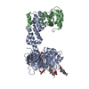
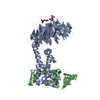
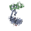
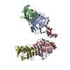
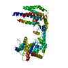
| Title | Three-dimensional structure of DNA sequence specificity (S) subunit of a type I restriction-modification enzyme and its functional implications | ||||||
 Components Components | Type I restriction-modification enzyme, S subunit | ||||||
 Keywords Keywords | HYDROLASE REGULATOR / Type I restriction modification enzyme / S-subunit / Structural genomics / PSI / Protein Structure Initiative / Berkeley Structural Genomics Center / BSGC | ||||||
| Function / homology |  Function and homology information Function and homology information | ||||||
| Biological species |   Methanocaldococcus jannaschii (archaea) Methanocaldococcus jannaschii (archaea) | ||||||
| Method |  X-RAY DIFFRACTION / SYNCHROTRON / SAD / Resolution: 2.4 Å X-RAY DIFFRACTION / SYNCHROTRON / SAD / Resolution: 2.4 Å | ||||||
 Authors Authors | Kim, J.S. / Degiovanni, A. / Jancarik, J. / Adams, P.D. / Yokota, H.A. / Kim, R. / Kim, S.H. / Berkeley Structural Genomics Center (BSGC) | ||||||
 Citation Citation | Journal: Proc.Natl.Acad.Sci.USA / Year: 2005 Title: Crystal structure of DNA sequence specificity subunit of a type I restriction-modification enzyme and its functional implications. Authors: Kim, J.S. / Degiovanni, A. / Jancarik, J. / Adams, P.D. / Yokota, H. / Kim, R. / Kim, S.H. | ||||||
| History |
|
- Structure visualization
| Structure viewer | Molecule: MolmilJmol/JSmol |
|---|
- Downloads & links
-Download
| PDBx/mmCIF format | 1yf2.cif.gz | 184.2 KB | Display | PDBx/mmCIF format |
|---|---|---|---|---|
| PDB format | pdb1yf2.ent.gz | 147.9 KB | Display | PDB format |
| PDBx/mmJSON format | 1yf2.json.gz | Tree view | PDBx/mmJSON format | |
| Others |  Other downloads Other downloads |
-Validation report
| Summary document | 1yf2_validation.pdf.gz | 424.3 KB | Display | wwPDB validaton report |
|---|---|---|---|---|
| Full document | 1yf2_full_validation.pdf.gz | 452.9 KB | Display | |
| Data in XML | 1yf2_validation.xml.gz | 37.1 KB | Display | |
| Data in CIF | 1yf2_validation.cif.gz | 52.2 KB | Display | |
| Arichive directory | https://data.pdbj.org/pub/pdb/validation_reports/yf/1yf2ftp://data.pdbj.org/pub/pdb/validation_reports/yf/1yf2 | HTTPS FTP |
-Related structure data
| Similar structure data | |
|---|---|
| Other databases |

-Links
 PDBj
PDBj- Assembly
| Deposited unit | 
| ||||||||
|---|---|---|---|---|---|---|---|---|---|
| 1 | 
| ||||||||
| 2 | 
| ||||||||
| Unit cell |
| ||||||||
| Details | The biological assembly is a monomer |
-Components
| #1: Protein | Mass: 48639.555 Da / Num. of mol.: 2 Source method: isolated from a genetically manipulated source Source: (gene. exp.) Methanocaldococcus jannaschii (archaea)Strain: DSM 2661 / Plasmid details: pET23a derivatized expression vector / Plasmid: pB3 / Species (production host): Escherichia coli / Production host:  #2: Water | ChemComp-HOH / |  Mass: 18.015 Da / Num. of mol.: 329 / Source method: isolated from a natural source / Formula: H2O Mass: 18.015 Da / Num. of mol.: 329 / Source method: isolated from a natural source / Formula: H2O |
|---|
-Experimental details
-Experiment
| Experiment | Method: X-RAY DIFFRACTION / Number of used crystals: 1 |
|---|
- Sample preparation
| Crystal | Density Matthews: 3.74 Å3/Da / Density % sol: 66.5 % |
|---|---|
| Crystal grow | Temperature: 298 K / Method: vapor diffusion, hanging drop / pH: 9.5 Details: PEG 3000, pH 9.5, VAPOR DIFFUSION, HANGING DROP, temperature 298.0K |
-Data collection
| Diffraction | Mean temperature: 100 K |
|---|---|
| Diffraction source | Source: SYNCHROTRON / Site: ALS  / Beamline: 8.2.2 / Wavelength: 1 Å / Beamline: 8.2.2 / Wavelength: 1 Å |
| Detector | Type: ADSC QUANTUM 4 / Detector: CCD / Date: Jul 12, 2004 / Details: mirrors |
| Radiation | Monochromator: graphite / Protocol: SINGLE WAVELENGTH / Monochromatic (M) / Laue (L): M / Scattering type: x-ray |
| Radiation wavelength | Wavelength: 1 Å / Relative weight: 1 |
| Reflection | Resolution: 2.21→50 Å / Num. all: 69051 / Num. obs: 59867 / Observed criterion σ(F): 1.2 / Observed criterion σ(I): 1.2 / Biso Wilson estimate: 49.2 Å2 |
| Reflection shell | Resolution: 2.21→2.29 Å / % possible all: 26.8 |
- Processing
| Software |
| ||||||||||||||||||||||||||||||||||||||||||||||||||||||||||||
|---|---|---|---|---|---|---|---|---|---|---|---|---|---|---|---|---|---|---|---|---|---|---|---|---|---|---|---|---|---|---|---|---|---|---|---|---|---|---|---|---|---|---|---|---|---|---|---|---|---|---|---|---|---|---|---|---|---|---|---|---|---|
| Refinement | Method to determine structure: SAD / Resolution: 2.4→48.52 Å / Rfactor Rfree error: 0.004 / Data cutoff high absF: 361731.16 / Data cutoff low absF: 0 / Isotropic thermal model: RESTRAINED / Cross valid method: THROUGHOUT / σ(F): 0 / Stereochemistry target values: Engh & Huber
| ||||||||||||||||||||||||||||||||||||||||||||||||||||||||||||
| Solvent computation | Solvent model: FLAT MODEL / Bsol: 60.6471 Å2 / ksol: 0.311875 e/Å3 | ||||||||||||||||||||||||||||||||||||||||||||||||||||||||||||
| Displacement parameters | Biso mean: 74.6 Å2
| ||||||||||||||||||||||||||||||||||||||||||||||||||||||||||||
| Refine analyze |
| ||||||||||||||||||||||||||||||||||||||||||||||||||||||||||||
| Refinement step | Cycle: LAST / Resolution: 2.4→48.52 Å
| ||||||||||||||||||||||||||||||||||||||||||||||||||||||||||||
| Refine LS restraints |
| ||||||||||||||||||||||||||||||||||||||||||||||||||||||||||||
| LS refinement shell | Resolution: 2.4→2.55 Å / Rfactor Rfree error: 0.015 / Total num. of bins used: 6
| ||||||||||||||||||||||||||||||||||||||||||||||||||||||||||||
| Xplor file |
|
