 Movie
Movie Controller

[English] 日本語
 Yorodumi
- PDB-1y6n: Crystal structure of Epstein-Barr virus IL-10 mutant (A87I) compl... -
+ Open data

- Basic information
| Entry | Database: PDB / ID: 1y6n | ||||||
|---|---|---|---|---|---|---|---|
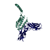
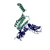
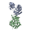
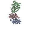
| Title | Crystal structure of Epstein-Barr virus IL-10 mutant (A87I) complexed with the soluble IL-10R1 chain | ||||||
 Components Components |
| ||||||
 Keywords Keywords |  IMMUNE SYSTEM / HELIX BUNDLE / RECEPTOR COMPLEX IMMUNE SYSTEM / HELIX BUNDLE / RECEPTOR COMPLEX | ||||||
| Function / homology |  Function and homology informationinterleukin-10 binding / interleukin-10 receptor activity / ubiquitin-dependent endocytosis / intestinal epithelial structure maintenance / regulation of synapse organization / Interleukin-10 signaling / regulation of cytokine production / negative regulation of autophagy / virus-mediated perturbation of host defense response / cytokine activity ...interleukin-10 binding / interleukin-10 receptor activity / ubiquitin-dependent endocytosis / intestinal epithelial structure maintenance / regulation of synapse organization / Interleukin-10 signaling / regulation of cytokine production / negative regulation of autophagy / virus-mediated perturbation of host defense response / cytokine activity / positive regulation of receptor signaling pathway via JAK-STAT / negative regulation of inflammatory response / cytokine-mediated signaling pathway / signaling receptor activity / response to lipopolysaccharide / immune response / apical plasma membrane / extracellular space / plasma membrane / cytosol Function and homology informationinterleukin-10 binding / interleukin-10 receptor activity / ubiquitin-dependent endocytosis / intestinal epithelial structure maintenance / regulation of synapse organization / Interleukin-10 signaling / regulation of cytokine production / negative regulation of autophagy / virus-mediated perturbation of host defense response / cytokine activity ...interleukin-10 binding / interleukin-10 receptor activity / ubiquitin-dependent endocytosis / intestinal epithelial structure maintenance / regulation of synapse organization / Interleukin-10 signaling / regulation of cytokine production / negative regulation of autophagy / virus-mediated perturbation of host defense response / cytokine activity / positive regulation of receptor signaling pathway via JAK-STAT / negative regulation of inflammatory response / cytokine-mediated signaling pathway / signaling receptor activity / response to lipopolysaccharide / immune response / apical plasma membrane / extracellular space / plasma membrane / cytosolSimilarity search - Function | ||||||
| Biological species |  Human herpesvirus 4 (Epstein-Barr virus) Human herpesvirus 4 (Epstein-Barr virus) Homo sapiens (human) Homo sapiens (human) | ||||||
| Method | X-RAY DIFFRACTION / SYNCHROTRON / MAD / Resolution: 2.7 Å | ||||||
 Authors Authors | Yoon, S.I. / Jones, B.C. / Logsdon, N.J. / Walter, M.R. | ||||||
 Citation Citation | Journal: Structure / Year: 2005 Title: Same structure, different function crystal structure of the Epstein-Barr virus IL-10 bound to the soluble IL-10R1 chain. Authors: Yoon, S.I. / Jones, B.C. / Logsdon, N.J. / Walter, M.R. | ||||||
| History |
|
- Structure visualization
| Structure viewer | Molecule: MolmilJmol/JSmol |
|---|
- Downloads & links
-Download
| PDBx/mmCIF format | 1y6n.cif.gz | 80.2 KB | Display | PDBx/mmCIF format |
|---|---|---|---|---|
| PDB format | pdb1y6n.ent.gz | 64.1 KB | Display | PDB format |
| PDBx/mmJSON format | 1y6n.json.gz | Tree view | PDBx/mmJSON format | |
| Others |  Other downloads Other downloads |
-Validation report
| Arichive directory | https://data.pdbj.org/pub/pdb/validation_reports/y6/1y6nftp://data.pdbj.org/pub/pdb/validation_reports/y6/1y6n | HTTPS FTP |
|---|
-Related structure data


-Links
 PDBj
PDBj


- Assembly
| Deposited unit | 
| ||||||||
|---|---|---|---|---|---|---|---|---|---|
| 1 | 
| ||||||||
| Unit cell |
| ||||||||
| Details | The second part of the biological assembly is generated by the two fold axis: x, x-y, -z. |
-Components
| #1: Protein | Mass: 17439.266 Da / Num. of mol.: 1 / Fragment: residues 26-170 / Mutation: A87I Source method: isolated from a genetically manipulated source Source: (gene. exp.) Human herpesvirus 4 (Epstein-Barr virus)Genus: Lymphocryptovirus / Strain: GD1 / Gene: BCRF1 / Plasmid: pET-32 / Production host:  Escherichia coli (E. coli) / Strain (production host): B834 (DE3) / References: UniProt: P03180 Escherichia coli (E. coli) / Strain (production host): B834 (DE3) / References: UniProt: P03180 |
|---|---|
| #2: Protein | Mass: 24506.551 Da / Num. of mol.: 1 / Fragment: Extracellular domain, residues 22-235 / Mutation: N29Q, N53Q, N89Q, N133Q, N156Q, N168Q Source method: isolated from a genetically manipulated source Source: (gene. exp.) Homo sapiens (human) / Gene: IL10RA, IL10R / Plasmid: pMTV5HIS / Cell line (production host): SCHNEIDER CELLS / Production host:  Drosophila melanogaster (fruit fly) / References: UniProt: Q13651 Drosophila melanogaster (fruit fly) / References: UniProt: Q13651 |
| #3: Water | ChemComp-HOH / Water Mass: 18.015 Da / Num. of mol.: 41 / Source method: isolated from a natural source / Formula: H2O Mass: 18.015 Da / Num. of mol.: 41 / Source method: isolated from a natural source / Formula: H2O |
-Experimental details
-Experiment
| Experiment | Method: X-RAY DIFFRACTION / Number of used crystals: 1 |
|---|
- Sample preparation
| Crystal | Density Matthews: 2.2 Å3/Da / Density % sol: 45 % |
|---|---|
| Crystal grow | Temperature: 298 K / Method: vapor diffusion, hanging drop / pH: 6.2 Details: PEG 6000, magnesium chloride, ADA, MPD, pH 6.2, VAPOR DIFFUSION, HANGING DROP, temperature 298K |
-Data collection
| Diffraction | Mean temperature: 100 K | |||||||||
|---|---|---|---|---|---|---|---|---|---|---|
| Diffraction source | Source: SYNCHROTRON / Site: SSRL  / Beamline: BL9-2 / Wavelength: 0.9252, 0.9794 / Beamline: BL9-2 / Wavelength: 0.9252, 0.9794 | |||||||||
| Detector | Type: ADSC QUANTUM 4 / Detector: CCD | |||||||||
| Radiation | Protocol: MAD / Monochromatic (M) / Laue (L): M / Scattering type: x-ray | |||||||||
| Radiation wavelength |
| |||||||||
| Reflection | Resolution: 2.7→60 Å / Num. all: 10500 / Num. obs: 10500 / % possible obs: 97.6 % / Observed criterion σ(F): 0 / Observed criterion σ(I): 0 / Redundancy: 4.3 % / Biso Wilson estimate: 57 Å2 / Rmerge(I) obs: 0.059 | |||||||||
| Reflection shell | Resolution: 2.7→2.8 Å / Rmerge(I) obs: 0.245 / % possible all: 85.1 |
- Processing
| Software |
| ||||||||||||||||||||
|---|---|---|---|---|---|---|---|---|---|---|---|---|---|---|---|---|---|---|---|---|---|
| Refinement | Method to determine structure: MAD / Resolution: 2.7→25 Å / σ(F): 0 / Stereochemistry target values: Engh & Huber
| ||||||||||||||||||||
| Refine analyze | Luzzati coordinate error free: 0.43 Å | ||||||||||||||||||||
| Refinement step | Cycle: LAST / Resolution: 2.7→25 Å
| ||||||||||||||||||||
| Refine LS restraints |
| ||||||||||||||||||||
| LS refinement shell | Resolution: 2.7→2.8 Å
|
