 Movie
Movie Controller

[English] 日本語
 Yorodumi
- PDB-1y67: Crystal Structure of Manganese Superoxide Dismutase from Deinococ... -
+ Open data

- Basic information
| Entry | Database: PDB / ID: 1y67 | ||||||
|---|---|---|---|---|---|---|---|
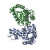
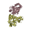
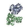
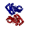
| Title | Crystal Structure of Manganese Superoxide Dismutase from Deinococcus radiodurans | ||||||
 Components Components | Manganese Superoxide dismutase Superoxide dismutase Superoxide dismutase | ||||||
 Keywords Keywords | OXIDOREDUCTASE / SUPEROXIDE DISMUTASE / MANGANESE ENZYME / METALLOPROTEIN | ||||||
| Function / homology |  Function and homology informationsuperoxide dismutase / superoxide dismutase activity / metal ion binding Function and homology informationsuperoxide dismutase / superoxide dismutase activity / metal ion bindingSimilarity search - Function | ||||||
| Biological species |  Deinococcus radiodurans (radioresistant) Deinococcus radiodurans (radioresistant) | ||||||
| Method | X-RAY DIFFRACTION / SYNCHROTRON / MOLECULAR REPLACEMENT / Resolution: 1.853 Å | ||||||
 Authors Authors | Chan, S. / Tanaka, S. / Sawaya, M.R. / Perry, L.J. | ||||||
 Citation Citation | Journal: To be Published Title: Crystal Structure of Manganese Superoxide Dismutase from Deinococcus radiodurans Authors: Chan, S. / Tanaka, S. / Sawaya, M.R. / Perry, L.J. #1: Journal: Acta Crystallogr.,Sect.D / Year: 1997Title: Refinement of Macromolecular Structures by the Maximum-Likelihood Method Authors: Murshudov, G.N. | ||||||
| History |
|
- Structure visualization
| Structure viewer | Molecule: MolmilJmol/JSmol |
|---|
- Downloads & links
-Download
| PDBx/mmCIF format | 1y67.cif.gz | 179.1 KB | Display | PDBx/mmCIF format |
|---|---|---|---|---|
| PDB format | pdb1y67.ent.gz | 141.6 KB | Display | PDB format |
| PDBx/mmJSON format | 1y67.json.gz | Tree view | PDBx/mmJSON format | |
| Others |  Other downloads Other downloads |
-Validation report
| Arichive directory | https://data.pdbj.org/pub/pdb/validation_reports/y6/1y67ftp://data.pdbj.org/pub/pdb/validation_reports/y6/1y67 | HTTPS FTP |
|---|
-Related structure data
| Related structure data | 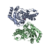 1vewS S: Starting model for refinement |
|---|---|
| Similar structure data |


-Links
 PDBj
PDBj
- Assembly
| Deposited unit | 
| ||||||||
|---|---|---|---|---|---|---|---|---|---|
| 1 | 
| ||||||||
| 2 | 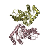
| ||||||||
| Unit cell |
| ||||||||
| Details | The biological assembly is a dimer formed from either chains A and B or chains C and D. |
-Components
| #1: Protein | Superoxide dismutase / MnSOD Mass: 25470.299 Da / Num. of mol.: 4 Source method: isolated from a genetically manipulated source Source: (gene. exp.) Deinococcus radiodurans (radioresistant)Gene: sodA / Plasmid: pET22b / Production host: Escherichia coli (E. coli) / Strain (production host): BL21-Gold (DE3) / References: UniProt: Q9RUV2, superoxide dismutase#2: Chemical | ChemComp-FE / Iron  Mass: 55.845 Da / Num. of mol.: 4 / Source method: obtained synthetically / Formula: Fe Mass: 55.845 Da / Num. of mol.: 4 / Source method: obtained synthetically / Formula: Fe#3: Water | ChemComp-HOH / | Water Mass: 18.015 Da / Num. of mol.: 396 / Source method: isolated from a natural source / Formula: H2O Mass: 18.015 Da / Num. of mol.: 396 / Source method: isolated from a natural source / Formula: H2O |
|---|
-Experimental details
-Experiment
| Experiment | Method: X-RAY DIFFRACTION / Number of used crystals: 1 |
|---|
- Sample preparation
| Crystal | Density Matthews: 2.43 Å3/Da / Density % sol: 49.04 % |
|---|---|
| Crystal grow | Temperature: 298 K / Method: vapor diffusion, sitting drop / pH: 7.1 Details: 200mM ammonium acetate (pH 7.1) 20%(w/v) PEG3350, 0.3M NaCl, 0.3M imidazole, 20mM Tris (pH 8.0), VAPOR DIFFUSION, SITTING DROP, temperature 298K |
-Data collection
| Diffraction | Mean temperature: 100 K |
|---|---|
| Diffraction source | Source: SYNCHROTRON / Site: ALS  / Beamline: 8.2.2 / Wavelength: 1.1 Å / Beamline: 8.2.2 / Wavelength: 1.1 Å |
| Detector | Type: ADSC QUANTUM 315 / Detector: CCD / Date: Apr 18, 2004 |
| Radiation | Monochromator: Double Crystal Si(111) / Protocol: SINGLE WAVELENGTH / Monochromatic (M) / Laue (L): M / Scattering type: x-ray |
| Radiation wavelength | Wavelength: 1.1 Å / Relative weight: 1 |
| Reflection | Resolution: 1.85→119.5 Å / Num. all: 74526 / Num. obs: 74526 / % possible obs: 99.8 % / Observed criterion σ(F): 0 / Observed criterion σ(I): 0 / Redundancy: 3.7 % / Biso Wilson estimate: 31.7 Å2 / Rsym value: 0.074 / Net I/σ(I): 15.4 |
| Reflection shell | Resolution: 1.85→1.92 Å / Redundancy: 3.5 % / Mean I/σ(I) obs: 2.6 / Num. unique all: 7437 / Rsym value: 0.493 / % possible all: 99.8 |
- Processing
| Software |
| |||||||||||||||||||||||||||||||||||||||||||||||||||||||||||||||||||||||||||||||||||||||||||||||||||||||||||||||||||||||||||||||||||||||||||||||||||
|---|---|---|---|---|---|---|---|---|---|---|---|---|---|---|---|---|---|---|---|---|---|---|---|---|---|---|---|---|---|---|---|---|---|---|---|---|---|---|---|---|---|---|---|---|---|---|---|---|---|---|---|---|---|---|---|---|---|---|---|---|---|---|---|---|---|---|---|---|---|---|---|---|---|---|---|---|---|---|---|---|---|---|---|---|---|---|---|---|---|---|---|---|---|---|---|---|---|---|---|---|---|---|---|---|---|---|---|---|---|---|---|---|---|---|---|---|---|---|---|---|---|---|---|---|---|---|---|---|---|---|---|---|---|---|---|---|---|---|---|---|---|---|---|---|---|---|---|---|
| Refinement | Method to determine structure: MOLECULAR REPLACEMENT Starting model: PDB ENTRY 1VEW Resolution: 1.853→119.523 Å / Cor.coef. Fo:Fc: 0.958 / Cor.coef. Fo:Fc free: 0.949 / WRfactor Rfree: 0.212 / WRfactor Rwork: 0.19 / Cross valid method: THROUGHOUT / σ(F): 0 / ESU R: 0.14 / ESU R Free: 0.121 / Stereochemistry target values: Engh & Huber / Details: HYDROGENS HAVE BEEN ADDED IN THE RIDING POSITIONS
| |||||||||||||||||||||||||||||||||||||||||||||||||||||||||||||||||||||||||||||||||||||||||||||||||||||||||||||||||||||||||||||||||||||||||||||||||||
| Solvent computation | Ion probe radii: 0.8 Å / Shrinkage radii: 0.8 Å / VDW probe radii: 1.2 Å / Solvent model: MASK BULK SOLVENT | |||||||||||||||||||||||||||||||||||||||||||||||||||||||||||||||||||||||||||||||||||||||||||||||||||||||||||||||||||||||||||||||||||||||||||||||||||
| Displacement parameters | Biso mean: 31.125 Å2
| |||||||||||||||||||||||||||||||||||||||||||||||||||||||||||||||||||||||||||||||||||||||||||||||||||||||||||||||||||||||||||||||||||||||||||||||||||
| Refine analyze | Luzzati coordinate error obs: 0.205 Å | |||||||||||||||||||||||||||||||||||||||||||||||||||||||||||||||||||||||||||||||||||||||||||||||||||||||||||||||||||||||||||||||||||||||||||||||||||
| Refinement step | Cycle: LAST / Resolution: 1.853→119.523 Å
| |||||||||||||||||||||||||||||||||||||||||||||||||||||||||||||||||||||||||||||||||||||||||||||||||||||||||||||||||||||||||||||||||||||||||||||||||||
| Refine LS restraints |
| |||||||||||||||||||||||||||||||||||||||||||||||||||||||||||||||||||||||||||||||||||||||||||||||||||||||||||||||||||||||||||||||||||||||||||||||||||
| LS refinement shell | Refine-ID: X-RAY DIFFRACTION / Total num. of bins used: 20
| |||||||||||||||||||||||||||||||||||||||||||||||||||||||||||||||||||||||||||||||||||||||||||||||||||||||||||||||||||||||||||||||||||||||||||||||||||
| Refinement TLS params. | Method: refined / Refine-ID: X-RAY DIFFRACTION
| |||||||||||||||||||||||||||||||||||||||||||||||||||||||||||||||||||||||||||||||||||||||||||||||||||||||||||||||||||||||||||||||||||||||||||||||||||
| Refinement TLS group | Refine-ID: X-RAY DIFFRACTION / Selection: ALL
|
