 Movie
Movie Controller

[English] 日本語
 Yorodumi
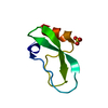
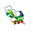
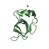
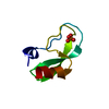
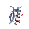
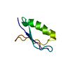
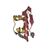
- PDB-1y62: A 2.4 crystal structure of conkunitzin-S1, a novel Kunitz-fold co... -
+ Open data

- Basic information
| Entry | Database: PDB / ID: 1y62 | ||||||
|---|---|---|---|---|---|---|---|
| Title | A 2.4 crystal structure of conkunitzin-S1, a novel Kunitz-fold cone snail neurotoxin. | ||||||
 Components Components | Conkunitzin-S1 | ||||||
 Keywords Keywords |  TOXIN / alpha helix / beta sheet / 310 helix / Kunitz fold TOXIN / alpha helix / beta sheet / 310 helix / Kunitz fold | ||||||
| Function / homology |  Function and homology information Function and homology informationpotassium channel regulator activity / serine-type endopeptidase inhibitor activity / toxin activity / extracellular regionSimilarity search - Function | ||||||
| Method | X-RAY DIFFRACTION / MOLECULAR REPLACEMENT / Resolution: 2.45 Å | ||||||
 Authors Authors | Dy, C.Y. / Buczek, P. / Horvath, M.P. | ||||||
 Citation Citation | Journal: Acta Crystallogr.,Sect.D / Year: 2006 Title: Structure of conkunitzin-S1, a neurotoxin and Kunitz-fold disulfide variant from cone snail. Authors: Dy, C.Y. / Buczek, P. / Imperial, J.S. / Bulaj, G. / Horvath, M.P. | ||||||
| History |
|
- Structure visualization
| Structure viewer | Molecule: MolmilJmol/JSmol |
|---|
- Downloads & links
-Download
| PDBx/mmCIF format | 1y62.cif.gz | 79.7 KB | Display | PDBx/mmCIF format |
|---|---|---|---|---|
| PDB format | pdb1y62.ent.gz | 61.4 KB | Display | PDB format |
| PDBx/mmJSON format | 1y62.json.gz | Tree view | PDBx/mmJSON format | |
| Others |  Other downloads Other downloads |
-Validation report
| Arichive directory | https://data.pdbj.org/pub/pdb/validation_reports/y6/1y62ftp://data.pdbj.org/pub/pdb/validation_reports/y6/1y62 | HTTPS FTP |
|---|
-Related structure data


-Links
 PDBj
PDBj


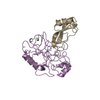
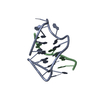
- Assembly
| Deposited unit | 
| ||||||||
|---|---|---|---|---|---|---|---|---|---|
| 1 | 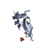
| ||||||||
| 2 | 
| ||||||||
| 3 | 
| ||||||||
| 4 | 
| ||||||||
| 5 | 
| ||||||||
| 6 | 
| ||||||||
| Unit cell |
| ||||||||
| Details | Each chain represents one biological unit. |
-Components
| #1: Protein | Mass: 6944.685 Da / Num. of mol.: 6 / Source method: obtained synthetically Details: The sequence of this peptide occurs naturally in Conus striatus (cone snail). This peptide was synthesized in two parts and subsequently joined through native chemical ligation. References: UniProt: P0C1X2*PLUS #2: Chemical | ChemComp-SO4 / Sulfate  Mass: 96.063 Da / Num. of mol.: 15 / Source method: obtained synthetically / Formula: SO4 Mass: 96.063 Da / Num. of mol.: 15 / Source method: obtained synthetically / Formula: SO4#3: Water | ChemComp-HOH / | Water Mass: 18.015 Da / Num. of mol.: 52 / Source method: isolated from a natural source / Formula: H2O Mass: 18.015 Da / Num. of mol.: 52 / Source method: isolated from a natural source / Formula: H2O |
|---|
-Experimental details
-Experiment
| Experiment | Method: X-RAY DIFFRACTION / Number of used crystals: 1 |
|---|
- Sample preparation
| Crystal | Density Matthews: 2.62 Å3/Da / Density % sol: 53.14 % |
|---|---|
| Crystal grow | Temperature: 291 K / Method: vapor diffusion, hanging drop / pH: 4 Details: PEG-400, ammonium sulfate, sodium azide, acetate, pH 4.0, VAPOR DIFFUSION, HANGING DROP, temperature 291K |
-Data collection
| Diffraction | Mean temperature: 100 K |
|---|---|
| Diffraction source | Source: ROTATING ANODE / Type: RIGAKU / Wavelength: 1.5418 |
| Detector | Type: Nonius Kappa CCD / Detector: CCD / Date: Aug 18, 2004 / Details: Nonius FR591 High brilliance |
| Radiation | Monochromator: Osmic MaxFlux (Green) / Protocol: SINGLE WAVELENGTH / Monochromatic (M) / Laue (L): M / Scattering type: x-ray |
| Radiation wavelength | Wavelength: 1.5418 Å / Relative weight: 1 |
| Reflection | Resolution: 2.45→20 Å / Num. all: 15511 / Num. obs: 15238 / % possible obs: 99 % / Observed criterion σ(F): 0 / Observed criterion σ(I): -3 / Redundancy: 4 % / Biso Wilson estimate: 28 Å2 / Rsym value: 0.098 |
| Reflection shell | Resolution: 2.45→2.54 Å / Redundancy: 3.8 % / Mean I/σ(I) obs: 3.8 / Num. unique all: 1498 / Rsym value: 0.365 / % possible all: 97.8 |
- Processing
| Software |
| ||||||||||||||||||||||||||||||||||||||||||||||||||||||||||||||||||||||||||||||||||||||||
|---|---|---|---|---|---|---|---|---|---|---|---|---|---|---|---|---|---|---|---|---|---|---|---|---|---|---|---|---|---|---|---|---|---|---|---|---|---|---|---|---|---|---|---|---|---|---|---|---|---|---|---|---|---|---|---|---|---|---|---|---|---|---|---|---|---|---|---|---|---|---|---|---|---|---|---|---|---|---|---|---|---|---|---|---|---|---|---|---|---|
| Refinement | Method to determine structure: MOLECULAR REPLACEMENT Starting model: ensemble of the kunitz domains in 1DTX,1KNT, 2PTC and 1TFX Resolution: 2.45→20 Å / Rfactor Rfree error: 0.007 / Data cutoff high absF: 122859.03 / Data cutoff low absF: 0 / Isotropic thermal model: RESTRAINED / Cross valid method: THROUGHOUT / σ(F): 0 / σ(I): -3 / Stereochemistry target values: Engh & Huber / Details: BULK SOLVENT MODEL USED
| ||||||||||||||||||||||||||||||||||||||||||||||||||||||||||||||||||||||||||||||||||||||||
| Solvent computation | Solvent model: FLAT MODEL / Bsol: 14.8714 Å2 / ksol: 0.4066 e/Å3 | ||||||||||||||||||||||||||||||||||||||||||||||||||||||||||||||||||||||||||||||||||||||||
| Displacement parameters | Biso mean: 23.9 Å2
| ||||||||||||||||||||||||||||||||||||||||||||||||||||||||||||||||||||||||||||||||||||||||
| Refine analyze |
| ||||||||||||||||||||||||||||||||||||||||||||||||||||||||||||||||||||||||||||||||||||||||
| Refinement step | Cycle: LAST / Resolution: 2.45→20 Å
| ||||||||||||||||||||||||||||||||||||||||||||||||||||||||||||||||||||||||||||||||||||||||
| Refine LS restraints |
| ||||||||||||||||||||||||||||||||||||||||||||||||||||||||||||||||||||||||||||||||||||||||
| LS refinement shell | Refine-ID: X-RAY DIFFRACTION / Total num. of bins used: 6
| ||||||||||||||||||||||||||||||||||||||||||||||||||||||||||||||||||||||||||||||||||||||||
| Xplor file |
|
