 Movie
Movie Controller

[English] 日本語
 Yorodumi
- PDB-1y60: Structure of the tetrahydromethanopterin dependent formaldehyde-a... -
+ Open data

- Basic information
| Entry | Database: PDB / ID: 1y60 | ||||||
|---|---|---|---|---|---|---|---|
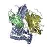
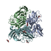
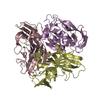
| Title | Structure of the tetrahydromethanopterin dependent formaldehyde-activating enzyme (Fae) from Methylobacterium extorquens AM1 with bound 5,10-methylene tetrahydromethanopterin | ||||||
 Components Components | Formaldehyde-activating enzyme fae | ||||||
 Keywords Keywords |  LYASE / Pentamer / beta-alpha-beta left handed crossover / TETRAHYDROMETHANOPTERIN-BINDING LYASE / Pentamer / beta-alpha-beta left handed crossover / TETRAHYDROMETHANOPTERIN-BINDING | ||||||
| Function / homology |  Function and homology information Function and homology information5,6,7,8-tetrahydromethanopterin hydro-lyase / carbon-nitrogen lyase activity / formaldehyde catabolic process / carbohydrate biosynthetic process / one-carbon metabolic process / cytoplasmSimilarity search - Function | ||||||
| Biological species |  Methylobacterium extorquens (bacteria) Methylobacterium extorquens (bacteria) | ||||||
| Method | X-RAY DIFFRACTION / SYNCHROTRON / MOLECULAR REPLACEMENT / Resolution: 1.9 Å | ||||||
 Authors Authors | Acharya, P. / Goenrich, M. / Hagemeier, C.H. / Demmer, U. / Vorholt, J.A. / Thauer, R.K. / Ermler, U. | ||||||
 Citation Citation | Journal: J.Biol.Chem. / Year: 2005 Title: How an enzyme binds the C1-carrier tetrahydromethanopterin: Structure of the tetrahydromethanopterin dependent formaldehyde-activating enzyme (Fae) from Methylobacterium extorquens AM1 Authors: Acharya, P. / Goenrich, M. / Hagemeier, C.H. / Demmer, U. / Vorholt, J.A. / Thauer, R.K. / Ermler, U. | ||||||
| History |
|
- Structure visualization
| Structure viewer | Molecule: MolmilJmol/JSmol |
|---|
- Downloads & links
-Download
| PDBx/mmCIF format | 1y60.cif.gz | 186.9 KB | Display | PDBx/mmCIF format |
|---|---|---|---|---|
| PDB format | pdb1y60.ent.gz | 146.6 KB | Display | PDB format |
| PDBx/mmJSON format | 1y60.json.gz | Tree view | PDBx/mmJSON format | |
| Others |  Other downloads Other downloads |
-Validation report
| Arichive directory | https://data.pdbj.org/pub/pdb/validation_reports/y6/1y60ftp://data.pdbj.org/pub/pdb/validation_reports/y6/1y60 | HTTPS FTP |
|---|
-Related structure data
| Related structure data |  1y5ySC S: Starting model for refinement C: citing same article ( |
|---|---|
| Similar structure data |
-Links
 PDBj
PDBj- Assembly
| Deposited unit | 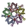
| ||||||||||
|---|---|---|---|---|---|---|---|---|---|---|---|
| 1 |
| ||||||||||
| Unit cell |
|
-Components
| #1: Protein | Mass: 17978.391 Da / Num. of mol.: 5 Source method: isolated from a genetically manipulated source Details: complexed with pterin-related cofactor / Source: (gene. exp.) Methylobacterium extorquens (bacteria) / Strain: AM1 / Gene: fae / Plasmid: pET17b / Production host: Escherichia coli (E. coli) / Strain (production host): BL21(DE3)pLysS / References: UniProt: Q9FA38#2: Chemical | ChemComp-H4M / 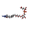  Mass: 788.693 Da / Num. of mol.: 5 / Source method: obtained synthetically / Formula: C31H45N6O16P Mass: 788.693 Da / Num. of mol.: 5 / Source method: obtained synthetically / Formula: C31H45N6O16P#3: Water | ChemComp-HOH / | Water Mass: 18.015 Da / Num. of mol.: 492 / Source method: isolated from a natural source / Formula: H2O Mass: 18.015 Da / Num. of mol.: 492 / Source method: isolated from a natural source / Formula: H2O |
|---|
-Experimental details
-Experiment
| Experiment | Method: X-RAY DIFFRACTION / Number of used crystals: 1 |
|---|
- Sample preparation
| Crystal | Density Matthews: 2 Å3/Da / Density % sol: 38 % |
|---|---|
| Crystal grow | Temperature: 281 K / Method: vapor diffusion, hanging drop / pH: 7.5 Details: 0.1 M HEPES/NaOH pH 7.5, 20% (w/v) polyethyleneglycol 10,000, 5 mM tetrahydromethanopterin (H4MPT), VAPOR DIFFUSION, HANGING DROP, temperature 281.0K |
-Data collection
| Diffraction | Mean temperature: 77 K |
|---|---|
| Diffraction source | Source: SYNCHROTRON / Site: ESRF  / Beamline: ID14-4 / Wavelength: 0.93927 Å / Beamline: ID14-4 / Wavelength: 0.93927 Å |
| Detector | Type: ADSC QUANTUM 4r / Detector: CCD / Date: Dec 7, 2003 |
| Radiation | Protocol: SINGLE WAVELENGTH / Monochromatic (M) / Laue (L): M / Scattering type: x-ray |
| Radiation wavelength | Wavelength: 0.93927 Å / Relative weight: 1 |
| Reflection | Resolution: 1.9→44.82 Å / Num. all: 59516 / Num. obs: 57191 / % possible obs: 93.5 % / Observed criterion σ(I): -3 / Redundancy: 3.7 % / Biso Wilson estimate: 20.9 Å2 / Rmerge(I) obs: 0.144 / Rsym value: 0.082 / Net I/σ(I): 12.15 |
| Reflection shell | Resolution: 1.9→2.01 Å / Redundancy: 3.2 % / Rmerge(I) obs: 0.5 / Mean I/σ(I) obs: 3.03 / Num. unique all: 8288 / Rsym value: 0.4 / % possible all: 76.1 |
- Processing
| Software |
| ||||||||||||||||||||||||||||||||||||
|---|---|---|---|---|---|---|---|---|---|---|---|---|---|---|---|---|---|---|---|---|---|---|---|---|---|---|---|---|---|---|---|---|---|---|---|---|---|
| Refinement | Method to determine structure: MOLECULAR REPLACEMENT Starting model: Formaldehyde-activating enzyme from Methylobacterium extorquens AM1 PDB ID 1Y5Y Resolution: 1.9→44.82 Å / Rfactor Rfree error: 0.004 / Data cutoff high absF: 108436.21 / Data cutoff low absF: 0 / Isotropic thermal model: RESTRAINED / Cross valid method: THROUGHOUT / σ(F): 0 / Stereochemistry target values: Engh & Huber
| ||||||||||||||||||||||||||||||||||||
| Solvent computation | Solvent model: FLAT MODEL / Bsol: 53.4963 Å2 / ksol: 0.390567 e/Å3 | ||||||||||||||||||||||||||||||||||||
| Displacement parameters | Biso mean: 29 Å2
| ||||||||||||||||||||||||||||||||||||
| Refine analyze |
| ||||||||||||||||||||||||||||||||||||
| Refinement step | Cycle: LAST / Resolution: 1.9→44.82 Å
| ||||||||||||||||||||||||||||||||||||
| Refine LS restraints |
| ||||||||||||||||||||||||||||||||||||
| LS refinement shell | Resolution: 1.9→2.02 Å / Rfactor Rfree error: 0.019 / Total num. of bins used: 6
| ||||||||||||||||||||||||||||||||||||
| Xplor file |
|
