 Movie
Movie Controller

[English] 日本語
 Yorodumi
- PDB-1y5h: Crystal structure of truncated Se-Met Hypoxic Response Protein I ... -
+ Open data

- Basic information
| Entry | Database: PDB / ID: 1y5h | ||||||
|---|---|---|---|---|---|---|---|
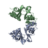
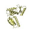
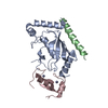
| Title | Crystal structure of truncated Se-Met Hypoxic Response Protein I (HRPI) | ||||||
 Components Components | hypothetical protein RV2626C Hypothesis Hypothesis | ||||||
 Keywords Keywords | UNKNOWN FUNCTION / CBS domain | ||||||
| Function / homology |  Function and homology information Function and homology informationsymbiont-mediated perturbation of host immune response / response to host immune response / peptidoglycan-based cell wall / response to hypoxia / extracellular region / metal ion binding / plasma membrane / cytosolSimilarity search - Function | ||||||
| Biological species |   Mycobacterium tuberculosis (bacteria) Mycobacterium tuberculosis (bacteria) | ||||||
| Method | X-RAY DIFFRACTION / SYNCHROTRON / MAD / Resolution: 1.5 Å | ||||||
 Authors Authors | Sharpe, M.L. / Baker, E.N. / Lott, J.S. | ||||||
 Citation Citation | Journal: J.Mol.Biol. / Year: 2008 Title: The structure and unusual protein chemistry of hypoxic response protein 1, a latency antigen and highly expressed member of the DosR regulon in Mycobacterium tuberculosis Authors: Sharpe, M.L. / Gao, C. / Kendall, S.L. / Baker, E.N. / Lott, J.S. | ||||||
| History |
|
- Structure visualization
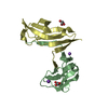
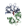
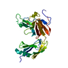
| Structure viewer | Molecule: MolmilJmol/JSmol |
|---|
- Downloads & links
-Download
| PDBx/mmCIF format | 1y5h.cif.gz | 56.6 KB | Display | PDBx/mmCIF format |
|---|---|---|---|---|
| PDB format | pdb1y5h.ent.gz | 44.1 KB | Display | PDB format |
| PDBx/mmJSON format | 1y5h.json.gz | Tree view | PDBx/mmJSON format | |
| Others |  Other downloads Other downloads |
-Validation report
| Arichive directory | https://data.pdbj.org/pub/pdb/validation_reports/y5/1y5hftp://data.pdbj.org/pub/pdb/validation_reports/y5/1y5h | HTTPS FTP |
|---|
-Related structure data

-Links
 PDBj
PDBj
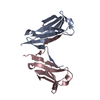
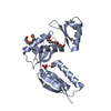
- Assembly
| Deposited unit | 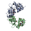
| ||||||||
|---|---|---|---|---|---|---|---|---|---|
| 1 |
| ||||||||
| Unit cell |
|
-Components
| #1: Protein | Hypothesis / hypoxic response protein I Mass: 14786.712 Da / Num. of mol.: 2 / Fragment: RESIDUES 1-127 Source method: isolated from a genetically manipulated source Source: (gene. exp.) Mycobacterium tuberculosis (bacteria) / Gene: Rv2626c / Plasmid: pET151 / Production host: Escherichia coli (E. coli) / Strain (production host): DL41 / References: UniProt: O06186, UniProt: P9WJA3*PLUS#2: Water | ChemComp-HOH / | Water Mass: 18.015 Da / Num. of mol.: 136 / Source method: isolated from a natural source / Formula: H2O Mass: 18.015 Da / Num. of mol.: 136 / Source method: isolated from a natural source / Formula: H2O |
|---|
-Experimental details
-Experiment
| Experiment | Method: X-RAY DIFFRACTION / Number of used crystals: 1 |
|---|
- Sample preparation
| Crystal | Density Matthews: 2 Å3/Da / Density % sol: 39.3 % / Description: the file contains Friedel pairs |
|---|---|
| Crystal grow | Temperature: 291 K / Method: vapor diffusion, sitting drop / pH: 9 Details: ammonium sulphate, boric acid/KOH, pH 9.0, VAPOR DIFFUSION, SITTING DROP, temperature 291K |
-Data collection
| Diffraction | Mean temperature: 100 K | ||||||||||||
|---|---|---|---|---|---|---|---|---|---|---|---|---|---|
| Diffraction source | Source: SYNCHROTRON / Site: ALS  / Beamline: 5.0.2 / Wavelength: 0.9793, 0.9796, 0.9537 / Beamline: 5.0.2 / Wavelength: 0.9793, 0.9796, 0.9537 | ||||||||||||
| Detector | Type: ADSC QUANTUM 315 / Detector: CCD / Date: Jul 3, 2004 / Details: Toroidal mirror | ||||||||||||
| Radiation | Monochromator: Double-crystal, Si(111) / Protocol: MAD / Monochromatic (M) / Laue (L): M / Scattering type: x-ray | ||||||||||||
| Radiation wavelength |
| ||||||||||||
| Reflection | Resolution: 1.5→50 Å / Num. all: 73443 / Num. obs: 66279 / % possible obs: 92.4 % / Redundancy: 6.2 % / Biso Wilson estimate: 22.5 Å2 / Limit h max: 51 / Limit h min: 0 / Limit k max: 27 / Limit k min: 0 / Limit l max: 49 / Limit l min: -52 / Observed criterion F max: 118610.22 / Observed criterion F min: 0.6 / Rsym value: 0.113 / Net I/σ(I): 20.97 | ||||||||||||
| Reflection shell | Resolution: 1.5→1.55 Å / Redundancy: 3.2 % / Mean I/σ(I) obs: 1.77 / Num. unique all: 2395 / Rsym value: 0.464 / % possible all: 63.7 |
- Processing
| Software |
| ||||||||||||||||||||||||||||||||||||||||||||||||||||||||||||||||||||||||||||||||||||||||||
|---|---|---|---|---|---|---|---|---|---|---|---|---|---|---|---|---|---|---|---|---|---|---|---|---|---|---|---|---|---|---|---|---|---|---|---|---|---|---|---|---|---|---|---|---|---|---|---|---|---|---|---|---|---|---|---|---|---|---|---|---|---|---|---|---|---|---|---|---|---|---|---|---|---|---|---|---|---|---|---|---|---|---|---|---|---|---|---|---|---|---|---|
| Refinement | Method to determine structure: MAD / Resolution: 1.5→38.06 Å / Rfactor Rfree error: 0.003 / Occupancy max: 1 / Occupancy min: 1 / Isotropic thermal model: anisotropic / Cross valid method: THROUGHOUT / Stereochemistry target values: Engh & Huber / Details: the file contains Friedel pairs
| ||||||||||||||||||||||||||||||||||||||||||||||||||||||||||||||||||||||||||||||||||||||||||
| Solvent computation | Solvent model: CNS bulk solvent model used / Bsol: 53.4993 Å2 / ksol: 0.385557 e/Å3 | ||||||||||||||||||||||||||||||||||||||||||||||||||||||||||||||||||||||||||||||||||||||||||
| Displacement parameters | Biso max: 56.94 Å2 / Biso mean: 26.8 Å2 / Biso min: 13.63 Å2
| ||||||||||||||||||||||||||||||||||||||||||||||||||||||||||||||||||||||||||||||||||||||||||
| Refine Biso |
| ||||||||||||||||||||||||||||||||||||||||||||||||||||||||||||||||||||||||||||||||||||||||||
| Refine analyze |
| ||||||||||||||||||||||||||||||||||||||||||||||||||||||||||||||||||||||||||||||||||||||||||
| Refinement step | Cycle: LAST / Resolution: 1.5→38.06 Å
| ||||||||||||||||||||||||||||||||||||||||||||||||||||||||||||||||||||||||||||||||||||||||||
| Refine LS restraints |
| ||||||||||||||||||||||||||||||||||||||||||||||||||||||||||||||||||||||||||||||||||||||||||
| LS refinement shell | Refine-ID: X-RAY DIFFRACTION / Total num. of bins used: 8
| ||||||||||||||||||||||||||||||||||||||||||||||||||||||||||||||||||||||||||||||||||||||||||
| Xplor file |
|
