 Movie
Movie Controller

[English] 日本語
 Yorodumi
- PDB-1xzo: Identification of a disulfide switch in BsSco, a member of the Sc... -
+ Open data

- Basic information
| Entry | Database: PDB / ID: 1xzo | |||||||||
|---|---|---|---|---|---|---|---|---|---|---|
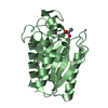
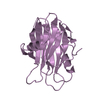
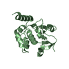
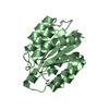
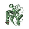
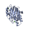
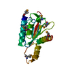
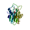
| Title | Identification of a disulfide switch in BsSco, a member of the Sco family of cytochrome c oxidase assembly proteins | |||||||||
 Components Components | Hypothetical protein ypmQ Hypothesis Hypothesis | |||||||||
 Keywords Keywords | METAL BINDING PROTEIN / thioredoxin-like fold / Structural Genomics / Montreal-Kingston Bacterial Structural Genomics Initiative / BSGI | |||||||||
| Function / homology |  Function and homology information Function and homology information | |||||||||
| Biological species |  Bacillus subtilis (bacteria) Bacillus subtilis (bacteria) | |||||||||
| Method | X-RAY DIFFRACTION / SYNCHROTRON / SAD / Resolution: 1.702 Å | |||||||||
 Authors Authors | Ye, Q. / Imriskova-Sosova, I. / Hill, B.C. / Jia, Z. / Montreal-Kingston Bacterial Structural Genomics Initiative (BSGI) | |||||||||
 Citation Citation | Journal: Biochemistry / Year: 2005 Title: Identification of a Disulfide Switch in BsSco, a Member of the Sco Family of Cytochrome c Oxidase Assembly Proteins Authors: Ye, Q. / Imriskova-Sosova, I. / Hill, B.C. / Jia, Z. | |||||||||
| History |
|
- Structure visualization
| Structure viewer | Molecule: MolmilJmol/JSmol |
|---|
- Downloads & links
-Download
| PDBx/mmCIF format | 1xzo.cif.gz | 95.8 KB | Display | PDBx/mmCIF format |
|---|---|---|---|---|
| PDB format | pdb1xzo.ent.gz | 72.1 KB | Display | PDB format |
| PDBx/mmJSON format | 1xzo.json.gz | Tree view | PDBx/mmJSON format | |
| Others |  Other downloads Other downloads |
-Validation report
| Arichive directory | https://data.pdbj.org/pub/pdb/validation_reports/xz/1xzoftp://data.pdbj.org/pub/pdb/validation_reports/xz/1xzo | HTTPS FTP |
|---|
-Related structure data
| Similar structure data | |
|---|---|
| Other databases |


-Links
 PDBj
PDBj- Assembly
| Deposited unit | 
| ||||||||
|---|---|---|---|---|---|---|---|---|---|
| 1 | 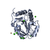
| ||||||||
| 2 | 
| ||||||||
| 3 | 
| ||||||||
| 4 |
| ||||||||
| Unit cell |
|
-Components
| #1: Protein | Hypothesis / BSSCO Mass: 19745.123 Da / Num. of mol.: 2 Source method: isolated from a genetically manipulated source Source: (gene. exp.) Bacillus subtilis (bacteria) / Strain: DH5f / Gene: ypmQ / Plasmid: PNM137 / Production host: Escherichia coli (E. coli) / References: UniProt: P54178#2: Chemical | ChemComp-CD /   Mass: 112.411 Da / Num. of mol.: 5 / Source method: obtained synthetically / Formula: Cd Mass: 112.411 Da / Num. of mol.: 5 / Source method: obtained synthetically / Formula: Cd#3: Chemical | ChemComp-CA /   Mass: 40.078 Da / Num. of mol.: 14 / Source method: obtained synthetically / Formula: Ca Mass: 40.078 Da / Num. of mol.: 14 / Source method: obtained synthetically / Formula: Ca#4: Water | ChemComp-HOH / | Water Mass: 18.015 Da / Num. of mol.: 388 / Source method: isolated from a natural source / Formula: H2O Mass: 18.015 Da / Num. of mol.: 388 / Source method: isolated from a natural source / Formula: H2O |
|---|
-Experimental details
-Experiment
| Experiment | Method: X-RAY DIFFRACTION / Number of used crystals: 1 |
|---|
- Sample preparation
| Crystal | Density Matthews: 3.12 Å3/Da / Density % sol: 60.3 % |
|---|---|
| Crystal grow | Temperature: 298 K / Method: vapor diffusion, hanging drop / pH: 6.5 Details: PEG5K MME, CdCl2, CaCl2, MES, pH 6.5, VAPOR DIFFUSION, HANGING DROP, temperature 298K |
-Data collection
| Diffraction | Mean temperature: 100 K |
|---|---|
| Diffraction source | Source: SYNCHROTRON / Site: NSLS  / Beamline: X12B / Wavelength: 0.9796 Å / Beamline: X12B / Wavelength: 0.9796 Å |
| Detector | Type: ADSC QUANTUM 4 / Detector: CCD / Date: Jun 27, 2003 |
| Radiation | Monochromator: Si(III) crystal / Protocol: SINGLE WAVELENGTH / Monochromatic (M) / Laue (L): M / Scattering type: x-ray |
| Radiation wavelength | Wavelength: 0.9796 Å / Relative weight: 1 |
| Reflection | Resolution: 1.7→64.55 Å / Num. all: 57817 / Num. obs: 54348 / Observed criterion σ(F): 0 / Observed criterion σ(I): 0 |
| Reflection shell | Resolution: 1.7→1.76 Å / % possible all: 0.94 |
- Processing
| Software |
| |||||||||||||||||||||||||||||||||||||||||||||||||||||||||||||||||||||||||||||||||||||||||||||||||||||||||
|---|---|---|---|---|---|---|---|---|---|---|---|---|---|---|---|---|---|---|---|---|---|---|---|---|---|---|---|---|---|---|---|---|---|---|---|---|---|---|---|---|---|---|---|---|---|---|---|---|---|---|---|---|---|---|---|---|---|---|---|---|---|---|---|---|---|---|---|---|---|---|---|---|---|---|---|---|---|---|---|---|---|---|---|---|---|---|---|---|---|---|---|---|---|---|---|---|---|---|---|---|---|---|---|---|---|---|
| Refinement | Method to determine structure: SAD / Resolution: 1.702→37.2 Å / Cor.coef. Fo:Fc: 0.933 / Cor.coef. Fo:Fc free: 0.9 / SU B: 3.079 / SU ML: 0.099 / Cross valid method: THROUGHOUT / σ(F): 0 / ESU R: 0.12 / ESU R Free: 0.126 / Stereochemistry target values: MAXIMUM LIKELIHOOD / Details: HYDROGENS HAVE BEEN ADDED IN THE RIDING POSITIONS
| |||||||||||||||||||||||||||||||||||||||||||||||||||||||||||||||||||||||||||||||||||||||||||||||||||||||||
| Solvent computation | Ion probe radii: 0.8 Å / Shrinkage radii: 0.8 Å / VDW probe radii: 1.4 Å / Solvent model: BABINET MODEL WITH MASK | |||||||||||||||||||||||||||||||||||||||||||||||||||||||||||||||||||||||||||||||||||||||||||||||||||||||||
| Displacement parameters | Biso mean: 26.669 Å2
| |||||||||||||||||||||||||||||||||||||||||||||||||||||||||||||||||||||||||||||||||||||||||||||||||||||||||
| Refinement step | Cycle: LAST / Resolution: 1.702→37.2 Å
| |||||||||||||||||||||||||||||||||||||||||||||||||||||||||||||||||||||||||||||||||||||||||||||||||||||||||
| Refine LS restraints |
| |||||||||||||||||||||||||||||||||||||||||||||||||||||||||||||||||||||||||||||||||||||||||||||||||||||||||
| LS refinement shell | Resolution: 1.702→1.746 Å / Total num. of bins used: 20 /
|
