 Movie
Movie Controller

[English] 日本語
 Yorodumi
- PDB-1xw4: Crystal Structure of Human Sulfiredoxin (Srx) in Complex with ADP -
+ Open data

- Basic information
| Entry | Database: PDB / ID: 1xw4 | ||||||
|---|---|---|---|---|---|---|---|
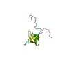
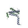
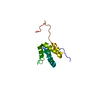
| Title | Crystal Structure of Human Sulfiredoxin (Srx) in Complex with ADP | ||||||
 Components Components | Sulfiredoxin | ||||||
 Keywords Keywords | OXIDOREDUCTASE / sulfiredoxin / cysteine / sulfinic acid / peroxiredoxin / sulphiredoxin | ||||||
| Function / homology |  Function and homology informationsulfiredoxin / oxidoreductase activity, acting on a sulfur group of donors / sulfiredoxin activity / NFE2L2 regulating anti-oxidant/detoxification enzymes / cellular response to oxidative stress / response to oxidative stress / endoplasmic reticulum membrane / ATP binding / cytosol / cytoplasm Function and homology informationsulfiredoxin / oxidoreductase activity, acting on a sulfur group of donors / sulfiredoxin activity / NFE2L2 regulating anti-oxidant/detoxification enzymes / cellular response to oxidative stress / response to oxidative stress / endoplasmic reticulum membrane / ATP binding / cytosol / cytoplasmSimilarity search - Function | ||||||
| Biological species |  Homo sapiens (human) Homo sapiens (human) | ||||||
| Method | X-RAY DIFFRACTION / MOLECULAR REPLACEMENT / Resolution: 2 Å | ||||||
 Authors Authors | Murray, M.S. / Jonsson, T.J. / Johnson, L.C. / Poole, L.B. / Lowther, W.T. | ||||||
 Citation Citation | Journal: Biochemistry / Year: 2005 Title: Structural basis for the retroreduction of inactivated peroxiredoxins by human sulfiredoxin. Authors: Jonsson, T.J. / Murray, M.S. / Johnson, L.C. / Poole, L.B. / Lowther, W.T. | ||||||
| History |
|
- Structure visualization
| Structure viewer | Molecule: MolmilJmol/JSmol |
|---|
- Downloads & links
-Download
| PDBx/mmCIF format | 1xw4.cif.gz | 38 KB | Display | PDBx/mmCIF format |
|---|---|---|---|---|
| PDB format | pdb1xw4.ent.gz | 24.6 KB | Display | PDB format |
| PDBx/mmJSON format | 1xw4.json.gz | Tree view | PDBx/mmJSON format | |
| Others |  Other downloads Other downloads |
-Validation report
| Arichive directory | https://data.pdbj.org/pub/pdb/validation_reports/xw/1xw4ftp://data.pdbj.org/pub/pdb/validation_reports/xw/1xw4 | HTTPS FTP |
|---|
-Related structure data
| Related structure data | 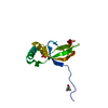 1xw3SC S: Starting model for refinement C: citing same article ( |
|---|---|
| Similar structure data |


-Links
 PDBj
PDBj- Assembly
| Deposited unit | 
| ||||||||
|---|---|---|---|---|---|---|---|---|---|
| 1 |
| ||||||||
| Unit cell |
|
-Components
| #1: Protein | Mass: 12066.685 Da / Num. of mol.: 1 / Mutation: residues 32-137 Source method: isolated from a genetically manipulated source Source: (gene. exp.) Homo sapiens (human) / Plasmid: pet19 / Production host:  Escherichia coli (E. coli) / Strain (production host): C41(DE3) / References: UniProt: Q9BYN0 Escherichia coli (E. coli) / Strain (production host): C41(DE3) / References: UniProt: Q9BYN0 |
|---|---|
| #2: Chemical | ChemComp-ADP / Adenosine diphosphate  Mass: 427.201 Da / Num. of mol.: 1 / Source method: obtained synthetically / Formula: C10H15N5O10P2 / Comment: ADP, energy-carrying molecule*YM Mass: 427.201 Da / Num. of mol.: 1 / Source method: obtained synthetically / Formula: C10H15N5O10P2 / Comment: ADP, energy-carrying molecule*YM |
| #3: Water | ChemComp-HOH / Water Mass: 18.015 Da / Num. of mol.: 71 / Source method: isolated from a natural source / Formula: H2O Mass: 18.015 Da / Num. of mol.: 71 / Source method: isolated from a natural source / Formula: H2O |
-Experimental details
-Experiment
| Experiment | Method: X-RAY DIFFRACTION / Number of used crystals: 1 |
|---|
- Sample preparation
| Crystal | Density Matthews: 2.9 Å3/Da / Density % sol: 56.7 % |
|---|---|
| Crystal grow | Temperature: 293 K / pH: 7.5 Details: MPD, pH 7.5, VAPOR DIFFUSION, HANGING DROP, temperature 293K, pH 7.50 |
-Data collection
| Diffraction | Mean temperature: 100 K |
|---|---|
| Diffraction source | Source: ROTATING ANODE / Type: RIGAKU MICROMAX-007 / Wavelength: 1.5418 |
| Detector | Type: RIGAKU / Detector: IMAGE PLATE / Date: Jul 9, 2004 / Details: CONFOCAL BLUE MAX-FLUX |
| Radiation | Protocol: SINGLE WAVELENGTH / Monochromatic (M) / Laue (L): M / Scattering type: x-ray |
| Radiation wavelength | Wavelength: 1.5418 Å / Relative weight: 1 |
| Reflection | Resolution: 2→29.64 Å / Num. obs: 9562 / % possible obs: 99.6 % / Observed criterion σ(I): 0 / Redundancy: 9.63 % / Rmerge(I) obs: 0.051 / Net I/σ(I): 30.9 |
| Reflection shell | Resolution: 2→2.07 Å / Redundancy: 5.03 % / Mean I/σ(I) obs: 4.4 / % possible all: 95.3 |
- Processing
| Software |
| ||||||||||||||||||||||||||||||||||||||||||||||||||||||||||||||||||||||||||||||||||||||||||||||||||||||||||||||||||||||||||||||||||||||||||||||||||||||||||||||||||||||||||
|---|---|---|---|---|---|---|---|---|---|---|---|---|---|---|---|---|---|---|---|---|---|---|---|---|---|---|---|---|---|---|---|---|---|---|---|---|---|---|---|---|---|---|---|---|---|---|---|---|---|---|---|---|---|---|---|---|---|---|---|---|---|---|---|---|---|---|---|---|---|---|---|---|---|---|---|---|---|---|---|---|---|---|---|---|---|---|---|---|---|---|---|---|---|---|---|---|---|---|---|---|---|---|---|---|---|---|---|---|---|---|---|---|---|---|---|---|---|---|---|---|---|---|---|---|---|---|---|---|---|---|---|---|---|---|---|---|---|---|---|---|---|---|---|---|---|---|---|---|---|---|---|---|---|---|---|---|---|---|---|---|---|---|---|---|---|---|---|---|---|---|---|
| Refinement | Method to determine structure: MOLECULAR REPLACEMENT Starting model: PDB ENTRY 1XW3 Resolution: 2→29.6 Å / Cor.coef. Fo:Fc: 0.944 / Cor.coef. Fo:Fc free: 0.917 / SU B: 4.501 / SU ML: 0.128 / Cross valid method: THROUGHOUT / ESU R: 0.187 / ESU R Free: 0.177 / Stereochemistry target values: MAXIMUM LIKELIHOOD / Details: HYDROGENS HAVE BEEN ADDED IN THE RIDING POSITIONS
| ||||||||||||||||||||||||||||||||||||||||||||||||||||||||||||||||||||||||||||||||||||||||||||||||||||||||||||||||||||||||||||||||||||||||||||||||||||||||||||||||||||||||||
| Solvent computation | Ion probe radii: 0.8 Å / Shrinkage radii: 0.8 Å / VDW probe radii: 1.2 Å / Solvent model: MASK | ||||||||||||||||||||||||||||||||||||||||||||||||||||||||||||||||||||||||||||||||||||||||||||||||||||||||||||||||||||||||||||||||||||||||||||||||||||||||||||||||||||||||||
| Displacement parameters | Biso mean: 27.872 Å2
| ||||||||||||||||||||||||||||||||||||||||||||||||||||||||||||||||||||||||||||||||||||||||||||||||||||||||||||||||||||||||||||||||||||||||||||||||||||||||||||||||||||||||||
| Refine analyze | Luzzati coordinate error obs: 0.36 Å / Luzzati sigma a obs: 0.23 Å | ||||||||||||||||||||||||||||||||||||||||||||||||||||||||||||||||||||||||||||||||||||||||||||||||||||||||||||||||||||||||||||||||||||||||||||||||||||||||||||||||||||||||||
| Refinement step | Cycle: LAST / Resolution: 2→29.6 Å
| ||||||||||||||||||||||||||||||||||||||||||||||||||||||||||||||||||||||||||||||||||||||||||||||||||||||||||||||||||||||||||||||||||||||||||||||||||||||||||||||||||||||||||
| Refine LS restraints |
| ||||||||||||||||||||||||||||||||||||||||||||||||||||||||||||||||||||||||||||||||||||||||||||||||||||||||||||||||||||||||||||||||||||||||||||||||||||||||||||||||||||||||||
| LS refinement shell | Resolution: 2.001→2.053 Å / Total num. of bins used: 20
|
