 Movie
Movie Controller


+ Open data

- Basic information
| Entry | Database: PDB / ID: 1xsm | ||||||
|---|---|---|---|---|---|---|---|
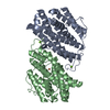
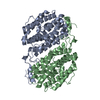
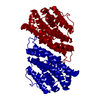
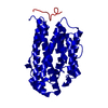
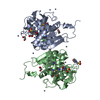
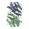
| Title | PROTEIN R2 OF RIBONUCLEOTIDE REDUCTASE FROM MOUSE | ||||||
 Components Components | RIBONUCLEOTIDE REDUCTASE R2 | ||||||
 Keywords Keywords |  OXIDOREDUCTASE / DNA REPLICATION / IRON OXIDOREDUCTASE / DNA REPLICATION / IRON | ||||||
| Function / homology |  Function and homology information Function and homology informationdeoxyribonucleotide metabolic process / Interconversion of nucleotide di- and triphosphates / ribonucleoside diphosphate metabolic process / 2'-deoxyribonucleotide biosynthetic process / ribonucleoside-diphosphate reductase complex / ribonucleoside-diphosphate reductase / ribonucleoside-diphosphate reductase activity, thioredoxin disulfide as acceptor / deoxyribonucleotide biosynthetic process / protein heterotetramerization / blastocyst development ...deoxyribonucleotide metabolic process / Interconversion of nucleotide di- and triphosphates / ribonucleoside diphosphate metabolic process / 2'-deoxyribonucleotide biosynthetic process / ribonucleoside-diphosphate reductase complex / ribonucleoside-diphosphate reductase / ribonucleoside-diphosphate reductase activity, thioredoxin disulfide as acceptor / deoxyribonucleotide biosynthetic process / protein heterotetramerization / blastocyst development / positive regulation of G1/S transition of mitotic cell cycle / ferric iron binding / nuclear envelope / protein homodimerization activity / identical protein binding / cytosolSimilarity search - Function | ||||||
| Biological species |  Mus musculus (house mouse) Mus musculus (house mouse) | ||||||
| Method | X-RAY DIFFRACTION / SYNCHROTRON / MOLECULAR REPLACEMENT / Resolution: 2.3 Å | ||||||
 Authors Authors | Kauppi, B. / Nielsen, B.N. / Ramaswamy, S. / Kjoller-Larsen, I. / Thelander, M. / Thelander, L. / Eklund, H. | ||||||
 Citation Citation | Journal: J.Mol.Biol. / Year: 1996 Title: The three-dimensional structure of mammalian ribonucleotide reductase protein R2 reveals a more-accessible iron-radical site than Escherichia coli R2. Authors: Kauppi, B. / Nielsen, B.B. / Ramaswamy, S. / Larsen, I.K. / Thelander, M. / Thelander, L. / Eklund, H. #1: Journal: J.Mol.Biol. / Year: 1993Title: Structure and Function of the Escherichia Coli Ribonucleotide Reductase Protein R2 Authors: Nordlund, P. / Eklund, H. | ||||||
| History |
|
- Structure visualization
| Structure viewer | Molecule: MolmilJmol/JSmol |
|---|
- Downloads & links
-Download
| PDBx/mmCIF format | 1xsm.cif.gz | 74.8 KB | Display | PDBx/mmCIF format |
|---|---|---|---|---|
| PDB format | pdb1xsm.ent.gz | 55.9 KB | Display | PDB format |
| PDBx/mmJSON format | 1xsm.json.gz | Tree view | PDBx/mmJSON format | |
| Others |  Other downloads Other downloads |
-Validation report
| Arichive directory | https://data.pdbj.org/pub/pdb/validation_reports/xs/1xsmftp://data.pdbj.org/pub/pdb/validation_reports/xs/1xsm | HTTPS FTP |
|---|
-Related structure data
| Related structure data |  1ribS S: Starting model for refinement |
|---|---|
| Similar structure data |


-Links
 PDBj
PDBj
- Assembly
| Deposited unit | 
| ||||||||
|---|---|---|---|---|---|---|---|---|---|
| 1 | 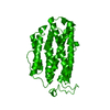
| ||||||||
| Unit cell |
|
-Components
| #1: Protein | Mass: 45149.547 Da / Num. of mol.: 1 Source method: isolated from a genetically manipulated source Source: (gene. exp.) Mus musculus (house mouse) / Description: T7-RNA POLYMERASE / Gene: NRDD / Plasmid: PET / Gene (production host): NRDD / Production host:  Escherichia coli (E. coli) Escherichia coli (E. coli)References: UniProt: P11157, ribonucleoside-diphosphate reductase |
|---|---|
| #2: Chemical | ChemComp-FE / Iron  Mass: 55.845 Da / Num. of mol.: 1 / Source method: obtained synthetically / Formula: Fe Mass: 55.845 Da / Num. of mol.: 1 / Source method: obtained synthetically / Formula: Fe |
| #3: Water | ChemComp-HOH / Water Mass: 18.015 Da / Num. of mol.: 90 / Source method: isolated from a natural source / Formula: H2O Mass: 18.015 Da / Num. of mol.: 90 / Source method: isolated from a natural source / Formula: H2O |
-Experimental details
-Experiment
| Experiment | Method: X-RAY DIFFRACTION / Number of used crystals: 9 |
|---|
- Sample preparation
| Crystal | Density Matthews: 2.14 Å3/Da / Density % sol: 40 % | ||||||||||||||||||||||||||||||||||||
|---|---|---|---|---|---|---|---|---|---|---|---|---|---|---|---|---|---|---|---|---|---|---|---|---|---|---|---|---|---|---|---|---|---|---|---|---|---|
| Crystal grow | pH: 4.7 Details: 1.0-1.2 M NACL, 0.1 M SODIUM ACETATE, PH 4.7, 0-4% PEG4000. | ||||||||||||||||||||||||||||||||||||
| Crystal | *PLUS | ||||||||||||||||||||||||||||||||||||
| Crystal grow | *PLUS Temperature: 20 ℃ / Method: vapor diffusion, hanging drop / Details: Nielsen, B.B., (1995) FEBS Letters, 373, 310. | ||||||||||||||||||||||||||||||||||||
| Components of the solutions | *PLUS
|
-Data collection
| Diffraction | Mean temperature: 277 K |
|---|---|
| Diffraction source | Source: SYNCHROTRON / Site: SRS  / Beamline: PX9.6 / Wavelength: 0.87 / Beamline: PX9.6 / Wavelength: 0.87 |
| Detector | Type: RIGAKU RAXIS IIC / Detector: IMAGE PLATE / Date: Feb 15, 1992 |
| Radiation | Monochromatic (M) / Laue (L): M / Scattering type: x-ray |
| Radiation wavelength | Wavelength: 0.87 Å / Relative weight: 1 |
| Reflection | Resolution: 2.3→25 Å / Num. obs: 17131 / % possible obs: 95.87 % / Observed criterion σ(I): 0 / Redundancy: 8.8 % / Rmerge(I) obs: 0.104 / Net I/σ(I): 17.6 |
| Reflection shell | Resolution: 2.29→2.38 Å / Redundancy: 4 % / Rmerge(I) obs: 0.283 / % possible all: 70.9 |
| Reflection | *PLUS % possible obs: 95.8 % / Num. measured all: 144719 |
| Reflection shell | *PLUS % possible obs: 70.9 % / Rmerge(I) obs: 0.3 |
- Processing
| Software |
| ||||||||||||||||||||||||||||||||||||||||||||||||||||||||||||
|---|---|---|---|---|---|---|---|---|---|---|---|---|---|---|---|---|---|---|---|---|---|---|---|---|---|---|---|---|---|---|---|---|---|---|---|---|---|---|---|---|---|---|---|---|---|---|---|---|---|---|---|---|---|---|---|---|---|---|---|---|---|
| Refinement | Method to determine structure: MOLECULAR REPLACEMENT Starting model: PDB ENTRY 1RIB Resolution: 2.3→25 Å / σ(F): 0
| ||||||||||||||||||||||||||||||||||||||||||||||||||||||||||||
| Displacement parameters | Biso mean: 38 Å2 | ||||||||||||||||||||||||||||||||||||||||||||||||||||||||||||
| Refinement step | Cycle: LAST / Resolution: 2.3→25 Å
| ||||||||||||||||||||||||||||||||||||||||||||||||||||||||||||
| Refine LS restraints |
| ||||||||||||||||||||||||||||||||||||||||||||||||||||||||||||
| Software | *PLUS Name: X-PLOR / Classification: refinement | ||||||||||||||||||||||||||||||||||||||||||||||||||||||||||||
| Refinement | *PLUS Rfactor Rfree: 0.25 | ||||||||||||||||||||||||||||||||||||||||||||||||||||||||||||
| Solvent computation | *PLUS | ||||||||||||||||||||||||||||||||||||||||||||||||||||||||||||
| Displacement parameters | *PLUS Biso mean: 35 Å2 | ||||||||||||||||||||||||||||||||||||||||||||||||||||||||||||
| Refine LS restraints | *PLUS Type: x_dihedral_angle_deg / Dev ideal: 28.6 |
