 Movie
Movie Controller

[English] 日本語
 Yorodumi
- PDB-1xsg: Solution structure of E.coli RNase P RNA P4 stem oligoribonucleot... -
+ Open data

- Basic information
| Entry | Database: PDB / ID: 1xsg | ||||||
|---|---|---|---|---|---|---|---|
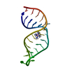
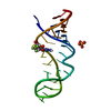
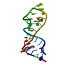
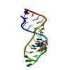
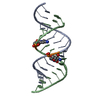
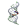
| Title | Solution structure of E.coli RNase P RNA P4 stem oligoribonucleotide, U69A mutation | ||||||
 Components Components | RNase P RNA P4 stem | ||||||
 Keywords Keywords |  RNA / Ribonuclease P RNA / Ribozyme / transfer RNA processing / P4 stem / U69A mutant / metal binding site RNA / Ribonuclease P RNA / Ribozyme / transfer RNA processing / P4 stem / U69A mutant / metal binding site | ||||||
| Function / homology | RNA / RNA (> 10) Function and homology information Function and homology information | ||||||
| Method | SOLUTION NMR / restrained molecular dynamics, simulated annealing | ||||||
| Model type details | minimized average | ||||||
 Authors Authors | Schmitz, M. | ||||||
 Citation Citation | Journal: Nucleic Acids Res. / Year: 2004 Title: Change of RNase P RNA function by single base mutation correlates with perturbation of metal ion binding in P4 as determined by NMR spectroscopy Authors: Schmitz, M. | ||||||
| History |
|
- Structure visualization
| Structure viewer | Molecule: MolmilJmol/JSmol |
|---|
- Downloads & links
-Download
| PDBx/mmCIF format | 1xsg.cif.gz | 23.3 KB | Display | PDBx/mmCIF format |
|---|---|---|---|---|
| PDB format | pdb1xsg.ent.gz | 16.9 KB | Display | PDB format |
| PDBx/mmJSON format | 1xsg.json.gz | Tree view | PDBx/mmJSON format | |
| Others |  Other downloads Other downloads |
-Validation report
| Arichive directory | https://data.pdbj.org/pub/pdb/validation_reports/xs/1xsgftp://data.pdbj.org/pub/pdb/validation_reports/xs/1xsg | HTTPS FTP |
|---|
-Related structure data


-Links
 PDBj
PDBj


- Assembly
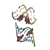
| Deposited unit | 
| |||||||||
|---|---|---|---|---|---|---|---|---|---|---|
| 1 |
| |||||||||
| NMR ensembles |
|
-Components
| #1: RNA chain | Mass: 8656.183 Da / Num. of mol.: 1 / Source method: obtained synthetically Details: enzymatically synthesized from DNA oligonucleotide template by T7 RNA polymerase |
|---|
-Experimental details
-Experiment
| Experiment | Method: SOLUTION NMR | ||||||||||||||||||||||||
|---|---|---|---|---|---|---|---|---|---|---|---|---|---|---|---|---|---|---|---|---|---|---|---|---|---|
| NMR experiment |
| ||||||||||||||||||||||||
| NMR details | Text: The structure was determined using standard 2D homonuclear techniques as well as 13C and 31P heteronuclear experiments performed at natural abundance |
- Sample preparation
| Details |
| |||||||||
|---|---|---|---|---|---|---|---|---|---|---|
| Sample conditions | Ionic strength: 100mM NaCl / pH: 6.4 / Pressure: ambient / Temperature: 288 K |
-NMR measurement
| Radiation | Protocol: SINGLE WAVELENGTH / Monochromatic (M) / Laue (L): M |
|---|---|
| Radiation wavelength | Relative weight: 1 |
| NMR spectrometer | Type: Bruker DMX / Manufacturer: Bruker / Model: DMX / Field strength: 500 MHz |
- Processing
| NMR software |
| ||||||||||||||||||||
|---|---|---|---|---|---|---|---|---|---|---|---|---|---|---|---|---|---|---|---|---|---|
| Refinement | Method: restrained molecular dynamics, simulated annealing / Software ordinal: 1 Details: The average structure is based on the superposition of 12 structures after refinement. The average RMS deviation between the ensemble and the average structure is 1.78 Angstrom. A total of ...Details: The average structure is based on the superposition of 12 structures after refinement. The average RMS deviation between the ensemble and the average structure is 1.78 Angstrom. A total of 290 NOE-derived distance constraints, 245 dihedral restraints and 48 distance restraints from hydrogen bonds were used in refinement. | ||||||||||||||||||||
| NMR representative | Selection criteria: minimized average structure | ||||||||||||||||||||
| NMR ensemble | Conformers submitted total number: 1 |
