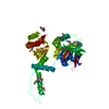
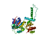
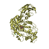
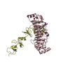
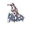
[English] 日本語
Yorodumi
- PDB-1xqs: Crystal structure of the HspBP1 core domain complexed with the fr... -

+
Open data


ID or keywords:

Loading...

-
Basic information

Entry
Database: PDB / ID: 1xqs
TitleCrystal structure of the HspBP1 core domain complexed with the fragment of Hsp70 ATPase domain
Components
  • HSPBP1 protein
  • Heat shock 70 kDa protein 1
KeywordsCHAPERONE / armadillo repeat / superhelical twist
Function / homology
Function and homology information


: / positive regulation of endoribonuclease activity / denatured protein binding / cellular heat acclimation / adenyl-nucleotide exchange factor activity / negative regulation of inclusion body assembly / Viral RNP Complexes in the Host Cell Nucleus / positive regulation of nucleotide-binding oligomerization domain containing 2 signaling pathway / : / death receptor agonist activity ...: / positive regulation of endoribonuclease activity / denatured protein binding / cellular heat acclimation / adenyl-nucleotide exchange factor activity / negative regulation of inclusion body assembly / Viral RNP Complexes in the Host Cell Nucleus / positive regulation of nucleotide-binding oligomerization domain containing 2 signaling pathway / : / death receptor agonist activity / C3HC4-type RING finger domain binding / ATP-dependent protein disaggregase activity / positive regulation of microtubule nucleation / molecular sequestering activity / misfolded protein binding / regulation of mitotic spindle assembly / negative regulation of mitochondrial outer membrane permeabilization involved in apoptotic signaling pathway / positive regulation of tumor necrosis factor-mediated signaling pathway / enzyme inhibitor activity / transcription regulator inhibitor activity / aggresome / lysosomal transport / cellular response to steroid hormone stimulus / mRNA catabolic process / chaperone cofactor-dependent protein refolding / regulation of protein ubiquitination / HSF1-dependent transactivation / response to unfolded protein / negative regulation of extrinsic apoptotic signaling pathway in absence of ligand / Regulation of HSF1-mediated heat shock response / chaperone-mediated protein complex assembly / Attenuation phase / cellular response to unfolded protein / negative regulation of endoplasmic reticulum stress-induced intrinsic apoptotic signaling pathway / ATP metabolic process / inclusion body / protein folding chaperone / vesicle-mediated transport / negative regulation of protein ubiquitination / heat shock protein binding / HSP90 chaperone cycle for steroid hormone receptors (SHR) in the presence of ligand / centriole / positive regulation of RNA splicing / positive regulation of erythrocyte differentiation / positive regulation of protein ubiquitination / AUF1 (hnRNP D0) binds and destabilizes mRNA / G protein-coupled receptor binding / positive regulation of interleukin-8 production / ATP-dependent protein folding chaperone / negative regulation of transforming growth factor beta receptor signaling pathway / PKR-mediated signaling / negative regulation of cell growth / histone deacetylase binding / transcription corepressor activity / disordered domain specific binding / unfolded protein binding / positive regulation of proteasomal ubiquitin-dependent protein catabolic process / protein folding / virus receptor activity / cellular response to oxidative stress / cellular response to heat / positive regulation of NF-kappaB transcription factor activity / protein refolding / blood microparticle / vesicle / ficolin-1-rich granule lumen / receptor ligand activity / protein stabilization / nuclear speck / cadherin binding / ribonucleoprotein complex / negative regulation of cell population proliferation / signaling receptor binding / focal adhesion / centrosome / ubiquitin protein ligase binding / Neutrophil degranulation / positive regulation of gene expression / negative regulation of apoptotic process / perinuclear region of cytoplasm / negative regulation of transcription by RNA polymerase II / enzyme binding / endoplasmic reticulum / ATP hydrolysis activity / protein-containing complex / mitochondrion / extracellular space / RNA binding / extracellular exosome / extracellular region / nucleoplasm / ATP binding / nucleus / plasma membrane / cytosol / cytoplasm
Similarity search - Function
Nucleotide exchange factor Fes1 / Nucleotide exchange factor Fes1 / Heat shock hsp70 proteins family signature 2. / Heat shock hsp70 proteins family signature 1. / Heat shock hsp70 proteins family signature 3. / Heat shock protein 70, conserved site / Heat shock protein 70kD, peptide-binding domain superfamily / Heat shock protein 70 family / Hsp70 protein / Heat shock protein 70kD, C-terminal domain superfamily ...Nucleotide exchange factor Fes1 / Nucleotide exchange factor Fes1 / Heat shock hsp70 proteins family signature 2. / Heat shock hsp70 proteins family signature 1. / Heat shock hsp70 proteins family signature 3. / Heat shock protein 70, conserved site / Heat shock protein 70kD, peptide-binding domain superfamily / Heat shock protein 70 family / Hsp70 protein / Heat shock protein 70kD, C-terminal domain superfamily / ATPase, substrate binding domain, subdomain 4 / Actin; Chain A, domain 4 / Leucine-rich Repeat Variant / Leucine-rich Repeat Variant / ATPase, nucleotide binding domain / ATPase, nucleotide binding domain / Armadillo-like helical / Alpha Horseshoe / Nucleotidyltransferase; domain 5 / Armadillo-type fold / Alpha-Beta Complex / 2-Layer Sandwich / Mainly Alpha / Alpha Beta
Similarity search - Domain/homology
ADENOSINE MONOPHOSPHATE / Heat shock 70 kDa protein 1B / Heat shock 70 kDa protein 1A / Hsp70-binding protein 1
Similarity search - Component
Biological speciesHomo sapiens (human)
MethodX-RAY DIFFRACTION / SYNCHROTRON / MOLECULAR REPLACEMENT / Resolution: 2.9 Å
AuthorsShomura, Y. / Dragovic, Z. / Chang, H.C. / Tzvetkov, N. / Young, J.C. / Brodsky, J.L. / Guerriero, V. / Hartl, F.U. / Bracher, A.
CitationJournal: Mol.Cell / Year: 2005
Title: Regulation of Hsp70 Function by HspBP1; Structural Analysis Reveals an Alternate Mechanism for Hsp70 Nucleotide Exchange
Authors: Shomura, Y. / Dragovic, Z. / Chang, H.C. / Tzvetkov, N. / Young, J.C. / Brodsky, J.L. / Guerriero, V. / Hartl, F.U. / Bracher, A.
History
DepositionOct 13, 2004Deposition site: RCSB / Processing site: PDBJ
Revision 1.0Mar 1, 2005Provider: repository / Type: Initial release
Revision 1.1Apr 30, 2008Group: Version format compliance
Revision 1.2Jul 13, 2011Group: Version format compliance
Revision 1.3Nov 10, 2021Group: Database references / Derived calculations / Category: database_2 / struct_ref_seq_dif / struct_site
Item: _database_2.pdbx_DOI / _database_2.pdbx_database_accession ..._database_2.pdbx_DOI / _database_2.pdbx_database_accession / _struct_ref_seq_dif.details / _struct_site.pdbx_auth_asym_id / _struct_site.pdbx_auth_comp_id / _struct_site.pdbx_auth_seq_id
Revision 1.4May 29, 2024Group: Data collection / Category: chem_comp_atom / chem_comp_bond

-
Structure visualization

Structure viewerMolecule:
MolmilJmol/JSmol

Downloads & links

-
Assembly

Deposited unit
A: HSPBP1 protein
B: HSPBP1 protein
C: Heat shock 70 kDa protein 1
D: Heat shock 70 kDa protein 1
hetero molecules


Theoretical massNumber of molelcules
Total (without water)106,1816
Polymers105,4874
Non-polymers6942
Water88349
1
A: HSPBP1 protein
C: Heat shock 70 kDa protein 1
hetero molecules


Theoretical massNumber of molelcules
Total (without water)53,0903
Polymers52,7432
Non-polymers3471
Water362
TypeNameSymmetry operationNumber
identity operation1_555x,y,z1
2
B: HSPBP1 protein
D: Heat shock 70 kDa protein 1
hetero molecules


Theoretical massNumber of molelcules
Total (without water)53,0903
Polymers52,7432
Non-polymers3471
Water362
TypeNameSymmetry operationNumber
identity operation1_555x,y,z1
Unit cell
Length a, b, c (Å)73.484, 94.809, 155.704
Angle α, β, γ (deg.)90.00, 90.00, 90.00
Int Tables number19
Space group name H-MP212121

-
Components

#1: Protein HSPBP1 protein


Mass: 31302.049 Da / Num. of mol.: 2 / Fragment: core domain (84-359) / Mutation: E88G
Source method: isolated from a genetically manipulated source
Source: (gene. exp.) Homo sapiens (human) / Plasmid: pPROExHTb / Production host: Escherichia coli (E. coli) / Strain (production host): BL21(DE3)codon plus RIL / References: UniProt: Q9NZL4
#2: Protein Heat shock 70 kDa protein 1 / Hsp70 / HSP70.1 / HSP70-1/HSP70-2


Mass: 21441.225 Da / Num. of mol.: 2 / Fragment: LOBE II OF ATPASE DOMAIN (184-371)
Source method: isolated from a genetically manipulated source
Source: (gene. exp.) Homo sapiens (human) / Plasmid: pPROExHTb / Production host: Escherichia coli (E. coli) / Strain (production host): BL21(DE3)codon plus RIL / References: UniProt: P08107, UniProt: P0DMV8*PLUS
#3: Chemical ChemComp-AMP / ADENOSINE MONOPHOSPHATE / Adenosine monophosphate


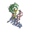
Mass: 347.221 Da / Num. of mol.: 2 / Source method: obtained synthetically / Formula: C10H14N5O7P / Comment: AMP*YM
#4: Water ChemComp-HOH / water / Water


Mass: 18.015 Da / Num. of mol.: 49 / Source method: isolated from a natural source / Formula: H2O

-
Experimental details

-
Experiment

ExperimentMethod: X-RAY DIFFRACTION / Number of used crystals: 1

-
Sample preparation

CrystalDensity Matthews: 2.57 Å3/Da / Density % sol: 64 %
Crystal growTemperature: 293 K / Method: vapor diffusion, hanging drop / pH: 7.5
Details: PEG8000, Potassium chloride, Magnesium chloride, AMP-PNP, DTT, HEPES KOH, pH 7.5, VAPOR DIFFUSION, HANGING DROP, temperature 293K

-
Data collection

DiffractionMean temperature: 100 K
Diffraction sourceSource: SYNCHROTRON / Site: ESRF / Beamline: ID14-2 / Wavelength: 0.933 Å
DetectorType: ADSC QUANTUM 4 / Detector: CCD / Date: Feb 20, 2004
RadiationMonochromator: diamond / Protocol: SINGLE WAVELENGTH / Monochromatic (M) / Laue (L): M / Scattering type: x-ray
Radiation wavelengthWavelength: 0.933 Å / Relative weight: 1
ReflectionResolution: 2.9→46.6 Å / Num. all: 24763 / Num. obs: 24763 / % possible obs: 99.8 % / Redundancy: 3.5 % / Biso Wilson estimate: 90.8 Å2 / Rsym value: 0.075 / Net I/σ(I): 6.5
Reflection shellResolution: 2.9→3.06 Å / Redundancy: 3.5 % / Mean I/σ(I) obs: 1.6 / Num. unique all: 3574 / Rsym value: 0.456 / % possible all: 100

-
Processing

Software
NameVersionClassification
MOSFLMdata reduction
SCALAdata scaling
BEASTmodel building
AMoREphasing
MOLREPphasing
CNS1refinement
CCP4(SCALA)data scaling
BEASTphasing
RefinementMethod to determine structure: MOLECULAR REPLACEMENT / Resolution: 2.9→46.6 Å / Isotropic thermal model: anisotropic / Cross valid method: THROUGHOUT / σ(F): 0 / Stereochemistry target values: Engh & Huber
RfactorNum. reflectionSelection details
Rfree0.296 1258 RANDOM
Rwork0.237 --
all-24763 -
obs-24730 -
Displacement parametersBiso mean: 51.4 Å2
Baniso -1Baniso -2Baniso -3
1--3.169 Å20 Å20 Å2
2--4.768 Å20 Å2
3---1.599 Å2
Refinement stepCycle: LAST / Resolution: 2.9→46.6 Å
ProteinNucleic acidLigandSolventTotal
Num. atoms6700 0 46 49 6795
Refine LS restraints
Refine-IDTypeDev ideal
X-RAY DIFFRACTIONc_bond_d0.007
X-RAY DIFFRACTIONc_angle_deg1.3
LS refinement shellResolution: 2.9→2.94 Å /
RfactorNum. reflection
Rfree0.367 51
Rwork0.457 -
obs-942

+
About Yorodumi

-
News

-
Feb 9, 2022. New format data for meta-information of EMDB entries

New format data for meta-information of EMDB entries

  • Version 3 of the EMDB header file is now the official format.
  • The previous official version 1.9 will be removed from the archive.

Related info.:EMDB header

External links:wwPDB to switch to version 3 of the EMDB data model

-
Aug 12, 2020. Covid-19 info

Covid-19 info

URL: https://pdbj.org/emnavi/covid19.php

New page: Covid-19 featured information page in EM Navigator.

Related info.:Covid-19 info / Mar 5, 2020. Novel coronavirus structure data

+
Mar 5, 2020. Novel coronavirus structure data

Novel coronavirus structure data

Related info.:Yorodumi Speices / Aug 12, 2020. Covid-19 info

External links:COVID-19 featured content - PDBj / Molecule of the Month (242):Coronavirus Proteases

+
Jan 31, 2019. EMDB accession codes are about to change! (news from PDBe EMDB page)

EMDB accession codes are about to change! (news from PDBe EMDB page)

  • The allocation of 4 digits for EMDB accession codes will soon come to an end. Whilst these codes will remain in use, new EMDB accession codes will include an additional digit and will expand incrementally as the available range of codes is exhausted. The current 4-digit format prefixed with “EMD-” (i.e. EMD-XXXX) will advance to a 5-digit format (i.e. EMD-XXXXX), and so on. It is currently estimated that the 4-digit codes will be depleted around Spring 2019, at which point the 5-digit format will come into force.
  • The EM Navigator/Yorodumi systems omit the EMD- prefix.

Related info.:Q: What is EMD? / ID/Accession-code notation in Yorodumi/EM Navigator

External links:EMDB Accession Codes are Changing Soon! / Contact to PDBj

+
Jul 12, 2017. Major update of PDB

Major update of PDB

  • wwPDB released updated PDB data conforming to the new PDBx/mmCIF dictionary.
  • This is a major update changing the version number from 4 to 5, and with Remediation, in which all the entries are updated.
  • In this update, many items about electron microscopy experimental information are reorganized (e.g. em_software).
  • Now, EM Navigator and Yorodumi are based on the updated data.

External links:wwPDB Remediation / Enriched Model Files Conforming to OneDep Data Standards Now Available in the PDB FTP Archive

-
Yorodumi

Thousand views of thousand structures

  • Yorodumi is a browser for structure data from EMDB, PDB, SASBDB, etc.
  • This page is also the successor to EM Navigator detail page, and also detail information page/front-end page for Omokage search.
  • The word "yorodu" (or yorozu) is an old Japanese word meaning "ten thousand". "mi" (miru) is to see.

Related info.:EMDB / PDB / SASBDB / Comparison of 3 databanks / Yorodumi Search / Aug 31, 2016. New EM Navigator & Yorodumi / Yorodumi Papers / Jmol/JSmol / Function and homology information / Changes in new EM Navigator and Yorodumi

Read more