 Movie
Movie Controller


+ Open data

- Basic information
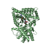
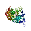
| Entry | Database: PDB / ID: 1xmc | ||||||
|---|---|---|---|---|---|---|---|
| Title | C323M mutant structure of mouse carnitine octanoyltransferase | ||||||
 Components Components | Peroxisomal carnitine O-octanoyltransferase | ||||||
 Keywords Keywords |  TRANSFERASE / carnitine / octanoyltransferase / hepes / mpd / mutant TRANSFERASE / carnitine / octanoyltransferase / hepes / mpd / mutant | ||||||
| Function / homology |  Function and homology information Function and homology informationBeta-oxidation of pristanoyl-CoA / carnitine O-octanoyltransferase / carnitine O-octanoyltransferase activity / medium-chain fatty acid metabolic process / carnitine metabolic process / Peroxisomal protein import / coenzyme A metabolic process / fatty acid beta-oxidation / fatty acid transport / fatty acid metabolic process ...Beta-oxidation of pristanoyl-CoA / carnitine O-octanoyltransferase / carnitine O-octanoyltransferase activity / medium-chain fatty acid metabolic process / carnitine metabolic process / Peroxisomal protein import / coenzyme A metabolic process / fatty acid beta-oxidation / fatty acid transport / fatty acid metabolic process / generation of precursor metabolites and energy / peroxisome / response to xenobiotic stimulus / intracellular membrane-bounded organelle / mitochondrionSimilarity search - Function | ||||||
| Biological species |  Mus musculus (house mouse) Mus musculus (house mouse) | ||||||
| Method | X-RAY DIFFRACTION / SYNCHROTRON / MOLECULAR REPLACEMENT / Resolution: 2 Å | ||||||
 Authors Authors | Jogl, G. / Hsiao, Y.S. / Tong, L. | ||||||
 Citation Citation | Journal: J.Biol.Chem. / Year: 2005 Title: Crystal structure of mouse carnitine octanoyltransferase and molecular determinants of substrate selectivity. Authors: Jogl, G. / Hsiao, Y.S. / Tong, L. | ||||||
| History |
|
- Structure visualization
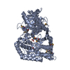
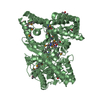
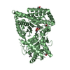
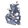
| Structure viewer | Molecule: MolmilJmol/JSmol |
|---|
- Downloads & links
-Download
| PDBx/mmCIF format | 1xmc.cif.gz | 259.5 KB | Display | PDBx/mmCIF format |
|---|---|---|---|---|
| PDB format | pdb1xmc.ent.gz | 209.4 KB | Display | PDB format |
| PDBx/mmJSON format | 1xmc.json.gz | Tree view | PDBx/mmJSON format | |
| Others |  Other downloads Other downloads |
-Validation report
| Arichive directory | https://data.pdbj.org/pub/pdb/validation_reports/xm/1xmcftp://data.pdbj.org/pub/pdb/validation_reports/xm/1xmc | HTTPS FTP |
|---|
-Related structure data


-Links
 PDBj
PDBj


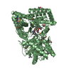
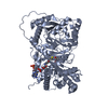
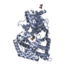
- Assembly
| Deposited unit | 
| ||||||||
|---|---|---|---|---|---|---|---|---|---|
| 1 | 
| ||||||||
| 2 | 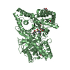
| ||||||||
| Unit cell |
|
-Components
| #1: Protein | Mass: 70385.430 Da / Num. of mol.: 2 / Mutation: C323M Source method: isolated from a genetically manipulated source Source: (gene. exp.) Mus musculus (house mouse) / Gene: Crot, Cot / Plasmid: pet28a / Production host:  Escherichia coli (E. coli) / Strain (production host): BL21 Star Escherichia coli (E. coli) / Strain (production host): BL21 StarReferences: UniProt: Q9DC50, carnitine O-octanoyltransferase#2: Chemical | HEPES  Mass: 238.305 Da / Num. of mol.: 2 / Source method: obtained synthetically / Formula: C8H18N2O4S / Comment: pH buffer*YM Mass: 238.305 Da / Num. of mol.: 2 / Source method: obtained synthetically / Formula: C8H18N2O4S / Comment: pH buffer*YM#3: Chemical | ChemComp-MPD / ( 2-Methyl-2,4-pentanediol  Mass: 118.174 Da / Num. of mol.: 7 / Source method: obtained synthetically / Formula: C6H14O2 / Comment: precipitant*YM Mass: 118.174 Da / Num. of mol.: 7 / Source method: obtained synthetically / Formula: C6H14O2 / Comment: precipitant*YM#4: Water | ChemComp-HOH / | Water Mass: 18.015 Da / Num. of mol.: 555 / Source method: isolated from a natural source / Formula: H2O Mass: 18.015 Da / Num. of mol.: 555 / Source method: isolated from a natural source / Formula: H2O |
|---|
-Experimental details
-Experiment
| Experiment | Method: X-RAY DIFFRACTION / Number of used crystals: 1 |
|---|
- Sample preparation
| Crystal | Density Matthews: 2.93 Å3/Da / Density % sol: 58 % |
|---|---|
| Crystal grow | Temperature: 277 K / Method: vapor diffusion, sitting drop / pH: 7.4 Details: 100mM Hepes, 62%v/v MPD, pH 7.4, VAPOR DIFFUSION, SITTING DROP, temperature 277K |
-Data collection
| Diffraction | Mean temperature: 100 K |
|---|---|
| Diffraction source | Source: SYNCHROTRON / Site: NSLS  / Beamline: X4A / Wavelength: 0.979 Å / Beamline: X4A / Wavelength: 0.979 Å |
| Detector | Type: ADSC QUANTUM 4 / Detector: CCD / Date: Apr 27, 2004 |
| Radiation | Protocol: SINGLE WAVELENGTH / Monochromatic (M) / Laue (L): M / Scattering type: x-ray |
| Radiation wavelength | Wavelength: 0.979 Å / Relative weight: 1 |
| Reflection | Resolution: 2→30 Å / Num. all: 99340 / Num. obs: 99340 / % possible obs: 92.5 % / Observed criterion σ(F): 0 / Observed criterion σ(I): 0 / Biso Wilson estimate: 13.4 Å2 |
| Reflection shell | Resolution: 2→2.07 Å / % possible all: 90.9 |
- Processing
| Software |
| |||||||||||||||||||||||||
|---|---|---|---|---|---|---|---|---|---|---|---|---|---|---|---|---|---|---|---|---|---|---|---|---|---|---|
| Refinement | Method to determine structure: MOLECULAR REPLACEMENT / Resolution: 2→26.75 Å / Rfactor Rfree error: 0.003 / Data cutoff high absF: 337446.55 / Data cutoff low absF: 0 / Isotropic thermal model: RESTRAINED / Cross valid method: THROUGHOUT / σ(F): 0 / Stereochemistry target values: Engh & Huber
| |||||||||||||||||||||||||
| Solvent computation | Solvent model: FLAT MODEL / Bsol: 50.4621 Å2 / ksol: 0.373225 e/Å3 | |||||||||||||||||||||||||
| Displacement parameters | Biso mean: 27 Å2
| |||||||||||||||||||||||||
| Refine analyze |
| |||||||||||||||||||||||||
| Refinement step | Cycle: LAST / Resolution: 2→26.75 Å
| |||||||||||||||||||||||||
| Refine LS restraints |
| |||||||||||||||||||||||||
| LS refinement shell | Resolution: 2→2.13 Å / Rfactor Rfree error: 0.007 / Total num. of bins used: 6
| |||||||||||||||||||||||||
| Xplor file |
|
