 Movie
Movie Controller

[English] 日本語
 Yorodumi
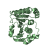
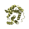
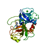
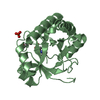
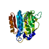
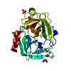
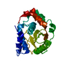
- PDB-1xm7: The Crystal Structure of the Protein of Unknown Function AQ665 fr... -
+ Open data

- Basic information
| Entry | Database: PDB / ID: 1xm7 | ||||||
|---|---|---|---|---|---|---|---|
| Title | The Crystal Structure of the Protein of Unknown Function AQ665 from Aquifex aeolicus | ||||||
 Components Components | hypothetical protein aq_1665 Hypothesis Hypothesis | ||||||
 Keywords Keywords | Structural genomics / unknown function / protein_aq1665 / protein structure initiative / midwest center for structural genomics / PSI / MCSG | ||||||
| Function / homology | Calcineurin-like phosphoesterase domain, lpxH-type / Calcineurin-like phosphoesterase superfamily domain / Metallo-dependent phosphatases / Purple Acid Phosphatase; chain A, domain 2 / Metallo-dependent phosphatase-like / 4-Layer Sandwich / Alpha Beta / Metallophos_2 domain-containing protein Function and homology information Function and homology information | ||||||
| Biological species |   Aquifex aeolicus (bacteria) Aquifex aeolicus (bacteria) | ||||||
| Method | X-RAY DIFFRACTION / SYNCHROTRON / MAD / Resolution: 2.4 Å | ||||||
 Authors Authors | Zhang, R. / Zhou, M. / Collart, F. / Joachimiak, A. / Midwest Center for Structural Genomics (MCSG) | ||||||
 Citation Citation | Journal: To be Published Title: The crystal structure of the hypothetical protein aq_1665 from Aquifex aeolicus Authors: Zhang, R. / Zhou, M. / Collart, F. / Joachimiak, A. / Midwest Center for Structural Genomics (MCSG) | ||||||
| History |
|
- Structure visualization
| Structure viewer | Molecule: MolmilJmol/JSmol |
|---|
- Downloads & links
-Download
| PDBx/mmCIF format | 1xm7.cif.gz | 85.9 KB | Display | PDBx/mmCIF format |
|---|---|---|---|---|
| PDB format | pdb1xm7.ent.gz | 70.9 KB | Display | PDB format |
| PDBx/mmJSON format | 1xm7.json.gz | Tree view | PDBx/mmJSON format | |
| Others |  Other downloads Other downloads |
-Validation report
| Arichive directory | https://data.pdbj.org/pub/pdb/validation_reports/xm/1xm7ftp://data.pdbj.org/pub/pdb/validation_reports/xm/1xm7 | HTTPS FTP |
|---|
-Related structure data
| Similar structure data | |
|---|---|
| Other databases |


-Links
 PDBj
PDBj
- Assembly
| Deposited unit | 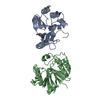
| ||||||||
|---|---|---|---|---|---|---|---|---|---|
| 1 | 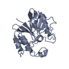
| ||||||||
| 2 | 
| ||||||||
| Unit cell |
| ||||||||
| Details | This protein existed as monomer.MolA and MolB represent two molecules in the asymmetric unit. |
-Components
| #1: Protein | Hypothesis Mass: 23687.150 Da / Num. of mol.: 2 Source method: isolated from a genetically manipulated source Source: (gene. exp.) Aquifex aeolicus (bacteria) / Strain: VF5 / Plasmid: PDM68 / Species (production host): Escherichia coli / Production host: Escherichia coli BL21 (bacteria) / Strain (production host): BL21 / References: UniProt: O67582#2: Water | ChemComp-HOH / | Water Mass: 18.015 Da / Num. of mol.: 70 / Source method: isolated from a natural source / Formula: H2O Mass: 18.015 Da / Num. of mol.: 70 / Source method: isolated from a natural source / Formula: H2O |
|---|
-Experimental details
-Experiment
| Experiment | Method: X-RAY DIFFRACTION / Number of used crystals: 1 |
|---|
- Sample preparation
| Crystal | Density Matthews: 3.05 Å3/Da / Density % sol: 58.1 % |
|---|---|
| Crystal grow | Temperature: 298 K / Method: vapor diffusion, sitting drop / pH: 7 Details: 0.1M Bis/Tris Propane, 2.8M Sodium Acetate, pH 7.0, VAPOR DIFFUSION, SITTING DROP, temperature 298K |
-Data collection
| Diffraction | Mean temperature: 100 K | ||||||||||||
|---|---|---|---|---|---|---|---|---|---|---|---|---|---|
| Diffraction source | Source: SYNCHROTRON / Site: APS  / Beamline: 19-ID / Wavelength: 0.9795,0.9798,0.9465 / Beamline: 19-ID / Wavelength: 0.9795,0.9798,0.9465 | ||||||||||||
| Detector | Type: SBC-2 / Detector: CCD / Date: Oct 26, 2003 / Details: mirrors | ||||||||||||
| Radiation | Monochromator: Si 111 channel / Protocol: MAD / Monochromatic (M) / Laue (L): M / Scattering type: x-ray | ||||||||||||
| Radiation wavelength |
| ||||||||||||
| Reflection | Resolution: 2.4→40 Å / Num. all: 22228 / Num. obs: 21851 / % possible obs: 98.3 % / Observed criterion σ(F): 2 / Observed criterion σ(I): 2 / Redundancy: 8.2 % / Biso Wilson estimate: 37.2 Å2 / Rmerge(I) obs: 0.106 / Net I/σ(I): 19.92 | ||||||||||||
| Reflection shell | Resolution: 2.4→2.49 Å / Redundancy: 6 % / Rmerge(I) obs: 0.695 / Mean I/σ(I) obs: 1.5 / Num. unique all: 2186 / % possible all: 87.2 |
- Processing
| Software |
| |||||||||||||||||||||||||
|---|---|---|---|---|---|---|---|---|---|---|---|---|---|---|---|---|---|---|---|---|---|---|---|---|---|---|
| Refinement | Method to determine structure: MAD / Resolution: 2.4→29.85 Å / Rfactor Rfree error: 0.006 / Data cutoff high absF: 262072.42 / Data cutoff low absF: 0 / Isotropic thermal model: RESTRAINED / Cross valid method: THROUGHOUT / σ(F): 0 / Stereochemistry target values: Engh & Huber / Details: Friedel pairs were used in the refinement.
| |||||||||||||||||||||||||
| Solvent computation | Solvent model: FLAT MODEL / Bsol: 39.4573 Å2 / ksol: 0.344659 e/Å3 | |||||||||||||||||||||||||
| Displacement parameters | Biso mean: 41.8 Å2
| |||||||||||||||||||||||||
| Refine analyze |
| |||||||||||||||||||||||||
| Refinement step | Cycle: LAST / Resolution: 2.4→29.85 Å
| |||||||||||||||||||||||||
| Refine LS restraints |
| |||||||||||||||||||||||||
| LS refinement shell | Resolution: 2.4→2.55 Å / Rfactor Rfree error: 0.022 / Total num. of bins used: 6
| |||||||||||||||||||||||||
| Xplor file |
|
