 Movie
Movie Controller

[English] 日本語
 Yorodumi
- PDB-1xhn: The crystal structure of Cellular Repressor of E1A-stimulated Gen... -
+ Open data

- Basic information
| Entry | Database: PDB / ID: 1xhn | ||||||
|---|---|---|---|---|---|---|---|
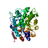
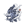
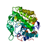
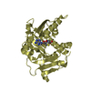
| Title | The crystal structure of Cellular Repressor of E1A-stimulated Genes (CREG) | ||||||
 Components Components | Cellular Repressor of E1A-stimulated Genes | ||||||
 Keywords Keywords | UNKNOWN FUNCTION /  beta-barrel beta-barrel | ||||||
| Function / homology |  Function and homology information Function and homology informationneutrophil degranulation / multicellular organism development / regulation of growth / transcription corepressor activity / azurophil granule lumen / Neutrophil degranulation / regulation of transcription by RNA polymerase II / extracellular space / extracellular exosome / extracellular regionSimilarity search - Function | ||||||
| Biological species |  Homo sapiens (human) Homo sapiens (human) | ||||||
| Method | X-RAY DIFFRACTION / SYNCHROTRON / MAD / Resolution: 1.95 Å | ||||||
 Authors Authors | Sacher, M. / Lunin, V.V. / Cygler, M. | ||||||
 Citation Citation | Journal: Proc.Natl.Acad.Sci.Usa / Year: 2005 Title: The crystal structure of CREG, a secreted glycoprotein involved in cellular growth and differentiation Authors: Sacher, M. / Di Bacco, A. / Lunin, V.V. / Ye, Z. / Wagner, J. / Gill, G. / Cygler, M. | ||||||
| History |
|
- Structure visualization
| Structure viewer | Molecule: MolmilJmol/JSmol |
|---|
- Downloads & links
-Download
| PDBx/mmCIF format | 1xhn.cif.gz | 163.3 KB | Display | PDBx/mmCIF format |
|---|---|---|---|---|
| PDB format | pdb1xhn.ent.gz | 137.3 KB | Display | PDB format |
| PDBx/mmJSON format | 1xhn.json.gz | Tree view | PDBx/mmJSON format | |
| Others |  Other downloads Other downloads |
-Validation report
| Arichive directory | https://data.pdbj.org/pub/pdb/validation_reports/xh/1xhnftp://data.pdbj.org/pub/pdb/validation_reports/xh/1xhn | HTTPS FTP |
|---|
-Related structure data
| Similar structure data |
|---|


-Links
 PDBj
PDBj- Assembly
| Deposited unit | 
| ||||||||
|---|---|---|---|---|---|---|---|---|---|
| 1 | 
| ||||||||
| 2 | 
| ||||||||
| Unit cell |
|
-Components
| #1: Protein | Mass: 20756.713 Da / Num. of mol.: 4 / Fragment: residues 13-184 Source method: isolated from a genetically manipulated source Source: (gene. exp.) Homo sapiens (human) / Production host:  Escherichia coli (E. coli) / References: UniProt: O75629 Escherichia coli (E. coli) / References: UniProt: O75629#2: Water | ChemComp-HOH / | Water Mass: 18.015 Da / Num. of mol.: 892 / Source method: isolated from a natural source / Formula: H2O Mass: 18.015 Da / Num. of mol.: 892 / Source method: isolated from a natural source / Formula: H2O |
|---|
-Experimental details
-Experiment
| Experiment | Method: X-RAY DIFFRACTION / Number of used crystals: 1 |
|---|
- Sample preparation
| Crystal | Density Matthews: 2.2 Å3/Da / Density % sol: 44.9 % |
|---|---|
| Crystal grow | Temperature: 298 K / Method: vapor diffusion, hanging drop / pH: 5 Details: Na acetate, PEG4000, ethylene glycole, pH 5.0, VAPOR DIFFUSION, HANGING DROP, temperature 298K |
-Data collection
| Diffraction | Mean temperature: 100 K | ||||||||||||
|---|---|---|---|---|---|---|---|---|---|---|---|---|---|
| Diffraction source | Source: SYNCHROTRON / Site: NSLS  / Beamline: X8C / Wavelength: 0.979502, 0.980005, 0.972318 / Beamline: X8C / Wavelength: 0.979502, 0.980005, 0.972318 | ||||||||||||
| Detector | Type: ADSC QUANTUM 4 / Detector: CCD / Date: Feb 6, 2004 | ||||||||||||
| Radiation | Protocol: MAD / Monochromatic (M) / Laue (L): M / Scattering type: x-ray | ||||||||||||
| Radiation wavelength |
| ||||||||||||
| Reflection | Resolution: 1.95→50 Å / Num. all: 51850 / Num. obs: 51850 / % possible obs: 100 % / Observed criterion σ(F): 2 / Observed criterion σ(I): 2 | ||||||||||||
| Reflection shell | Resolution: 1.95→2.02 Å / % possible all: 100 |
- Processing
| Software |
| ||||||||||||||||||||||||||||||||||||||||||||||||||||||||||||||||||||||
|---|---|---|---|---|---|---|---|---|---|---|---|---|---|---|---|---|---|---|---|---|---|---|---|---|---|---|---|---|---|---|---|---|---|---|---|---|---|---|---|---|---|---|---|---|---|---|---|---|---|---|---|---|---|---|---|---|---|---|---|---|---|---|---|---|---|---|---|---|---|---|---|
| Refinement | Method to determine structure: MAD / Resolution: 1.95→19.96 Å / Cor.coef. Fo:Fc: 0.955 / Cor.coef. Fo:Fc free: 0.919 / SU B: 3.698 / SU ML: 0.107 / Cross valid method: THROUGHOUT / σ(F): 0 / ESU R: 0.16 / ESU R Free: 0.149 / Stereochemistry target values: MAXIMUM LIKELIHOOD
| ||||||||||||||||||||||||||||||||||||||||||||||||||||||||||||||||||||||
| Solvent computation | Ion probe radii: 0.8 Å / Shrinkage radii: 0.8 Å / VDW probe radii: 1.4 Å / Solvent model: BABINET MODEL WITH MASK | ||||||||||||||||||||||||||||||||||||||||||||||||||||||||||||||||||||||
| Displacement parameters | Biso mean: 20.999 Å2
| ||||||||||||||||||||||||||||||||||||||||||||||||||||||||||||||||||||||
| Refinement step | Cycle: LAST / Resolution: 1.95→19.96 Å
| ||||||||||||||||||||||||||||||||||||||||||||||||||||||||||||||||||||||
| Refine LS restraints |
| ||||||||||||||||||||||||||||||||||||||||||||||||||||||||||||||||||||||
| LS refinement shell | Resolution: 1.952→2.002 Å / Total num. of bins used: 20 /
|
