 Movie
Movie Controller

[English] 日本語
 Yorodumi
- PDB-1xg5: Structure of human putative dehydrogenase MGC4172 in complex with NADP -
+ Open data

- Basic information
| Entry | Database: PDB / ID: 1xg5 | ||||||
|---|---|---|---|---|---|---|---|
| Title | Structure of human putative dehydrogenase MGC4172 in complex with NADP | ||||||
 Components Components | ARPG836 | ||||||
 Keywords Keywords |  OXIDOREDUCTASE / Short Chain Dehydrogenase / Human / SGC / structural genomics / Structural Genomics Consortium OXIDOREDUCTASE / Short Chain Dehydrogenase / Human / SGC / structural genomics / Structural Genomics Consortium | ||||||
| Function / homology |  Function and homology information Function and homology information3beta-hydroxysteroid 3-dehydrogenase / 3-keto sterol reductase activity / 17-beta-ketosteroid reductase activity / 17-beta-hydroxysteroid dehydrogenase (NAD+) activity / 17-beta-hydroxysteroid dehydrogenase (NADP+) activity / estrogen biosynthetic process / 17beta-estradiol 17-dehydrogenase / estradiol 17-beta-dehydrogenase [NAD(P)] activity / steroid biosynthetic process / nucleotide binding / extracellular regionSimilarity search - Function | ||||||
| Biological species |  Homo sapiens (human) Homo sapiens (human) | ||||||
| Method | X-RAY DIFFRACTION / SYNCHROTRON / MOLECULAR REPLACEMENT / Resolution: 1.53 Å | ||||||
 Authors Authors | Kavanagh, K. / Ng, S. / Sharma, S. / Vedadi, M. / von Delft, F. / Walker, J.R. / dhe Paganon, S. / Bray, J. / Oppermann, U. / Edwards, A. ...Kavanagh, K. / Ng, S. / Sharma, S. / Vedadi, M. / von Delft, F. / Walker, J.R. / dhe Paganon, S. / Bray, J. / Oppermann, U. / Edwards, A. / Arrowsmith, C. / Sundstrom, M. / Structural Genomics Consortium (SGC) | ||||||
 Citation Citation | Journal: To be Published Title: Structural Genomics Consortium: Structure of the putative human dehydrogenase MGC4172 Authors: Kavanagh, K. / Ng, S. / Sharma, S. / Vedadi, M. / von Delft, F. / Walker, J.R. / dhe Paganon, S. / Bray, J. / Oppermann, U. / Edwards, A. / Arrowsmith, C. / Sundstrom, M. | ||||||
| History |
|
- Structure visualization
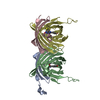
| Structure viewer | Molecule: MolmilJmol/JSmol |
|---|
- Downloads & links
-Download
| PDBx/mmCIF format | 1xg5.cif.gz | 227.6 KB | Display | PDBx/mmCIF format |
|---|---|---|---|---|
| PDB format | pdb1xg5.ent.gz | 180.7 KB | Display | PDB format |
| PDBx/mmJSON format | 1xg5.json.gz | Tree view | PDBx/mmJSON format | |
| Others |  Other downloads Other downloads |
-Validation report
| Arichive directory | https://data.pdbj.org/pub/pdb/validation_reports/xg/1xg5ftp://data.pdbj.org/pub/pdb/validation_reports/xg/1xg5 | HTTPS FTP |
|---|
-Related structure data
| Related structure data |  1edoS S: Starting model for refinement |
|---|---|
| Similar structure data |


-Links
 PDBj
PDBj


- Assembly
| Deposited unit | 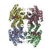
| ||||||||
|---|---|---|---|---|---|---|---|---|---|
| 1 |
| ||||||||
| 2 | 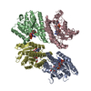
| ||||||||
| Unit cell |
| ||||||||
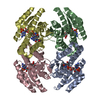
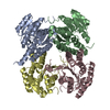
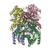
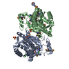
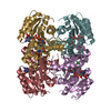
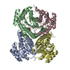
| Details | The biological assembly is a tetramer, contained in one asymmetric unit. |
-Components
| #1: Protein | Mass: 30466.518 Da / Num. of mol.: 4 Source method: isolated from a genetically manipulated source Source: (gene. exp.) Homo sapiens (human) / Gene: MGC4172 / Plasmid: p11 (pET11 derivative) / Production host:  Escherichia coli (E. coli) / Strain (production host): rosetta2 / References: UniProt: Q6UWP2 Escherichia coli (E. coli) / Strain (production host): rosetta2 / References: UniProt: Q6UWP2#2: Chemical | ChemComp-NAP / Nicotinamide adenine dinucleotide phosphate  Mass: 743.405 Da / Num. of mol.: 4 / Source method: obtained synthetically / Formula: C21H28N7O17P3 Mass: 743.405 Da / Num. of mol.: 4 / Source method: obtained synthetically / Formula: C21H28N7O17P3#3: Chemical | Acetic acid  Mass: 60.052 Da / Num. of mol.: 3 / Source method: obtained synthetically / Formula: C2H4O2 Mass: 60.052 Da / Num. of mol.: 3 / Source method: obtained synthetically / Formula: C2H4O2#4: Water | ChemComp-HOH / | Water Mass: 18.015 Da / Num. of mol.: 859 / Source method: isolated from a natural source / Formula: H2O Mass: 18.015 Da / Num. of mol.: 859 / Source method: isolated from a natural source / Formula: H2O |
|---|
-Experimental details
-Experiment
| Experiment | Method: X-RAY DIFFRACTION / Number of used crystals: 1 |
|---|
- Sample preparation
| Crystal | Density Matthews: 2 Å3/Da / Density % sol: 39 % |
|---|---|
| Crystal grow | Temperature: 293 K / Method: vapor diffusion, sitting drop / pH: 5.5 Details: 100mM Bis-Tris pH 5.5, 200mM Ammonium Acetate, 2% Glycerol (total 7%), 18% PEG 3350, 0.5mM TCEP, VAPOR DIFFUSION, SITTING DROP, temperature 293K |
-Data collection
| Diffraction | Mean temperature: 100 K |
|---|---|
| Diffraction source | Source: SYNCHROTRON / Site: APS  / Beamline: 17-ID / Wavelength: 1 Å / Beamline: 17-ID / Wavelength: 1 Å |
| Detector | Type: ADSC QUANTUM 4 / Detector: CCD / Date: Aug 6, 2004 |
| Radiation | Monochromator: Mirrors / Protocol: SINGLE WAVELENGTH / Monochromatic (M) / Laue (L): M / Scattering type: x-ray |
| Radiation wavelength | Wavelength: 1 Å / Relative weight: 1 |
| Reflection | Resolution: 1.53→41.56 Å / Num. all: 149996 / Num. obs: 145577 / % possible obs: 97.05 % / Observed criterion σ(F): 0 / Observed criterion σ(I): 0 / Redundancy: 5.3 % / Biso Wilson estimate: 14.99 Å2 / Rmerge(I) obs: 0.087 / Rsym value: 0.087 / Net I/σ(I): 13 |
| Reflection shell | Resolution: 1.53→1.57 Å / Redundancy: 1.6 % / Rmerge(I) obs: 0.558 / Mean I/σ(I) obs: 1.6 / Num. unique all: 8970 / Rsym value: 0.558 / % possible all: 81.2 |
- Processing
| Software |
| ||||||||||||||||||||||||||||||||||||||||||||||||||||||||||||||||||||||||||||||||||||||||||||||||||||||||||||||||||||||||||||||||||||||||||||||||||||||||||||||||
|---|---|---|---|---|---|---|---|---|---|---|---|---|---|---|---|---|---|---|---|---|---|---|---|---|---|---|---|---|---|---|---|---|---|---|---|---|---|---|---|---|---|---|---|---|---|---|---|---|---|---|---|---|---|---|---|---|---|---|---|---|---|---|---|---|---|---|---|---|---|---|---|---|---|---|---|---|---|---|---|---|---|---|---|---|---|---|---|---|---|---|---|---|---|---|---|---|---|---|---|---|---|---|---|---|---|---|---|---|---|---|---|---|---|---|---|---|---|---|---|---|---|---|---|---|---|---|---|---|---|---|---|---|---|---|---|---|---|---|---|---|---|---|---|---|---|---|---|---|---|---|---|---|---|---|---|---|---|---|---|---|---|
| Refinement | Method to determine structure: MOLECULAR REPLACEMENT Starting model: PDB entry 1EDO Resolution: 1.53→41.56 Å / Cor.coef. Fo:Fc: 0.969 / Cor.coef. Fo:Fc free: 0.949 / SU B: 3.004 / SU ML: 0.054 / TLS residual ADP flag: LIKELY RESIDUAL Isotropic thermal model: Individual isotropic thermal parameters Cross valid method: THROUGHOUT / σ(F): 0 / σ(I): 0 / ESU R: 0.071 / ESU R Free: 0.079 / Stereochemistry target values: MAXIMUM LIKELIHOOD Details: 1) HYDROGENS HAVE BEEN ADDED IN THE RIDING POSITIONS. 2) PROMINENT DIFFERENCE DENSITY AROUND RESIDUES A208, C208, D208 AND B151 WAS NOT INTERPRETABLE AND WAS LEFT UNMODELLED.
| ||||||||||||||||||||||||||||||||||||||||||||||||||||||||||||||||||||||||||||||||||||||||||||||||||||||||||||||||||||||||||||||||||||||||||||||||||||||||||||||||
| Solvent computation | Ion probe radii: 0.8 Å / Shrinkage radii: 0.8 Å / VDW probe radii: 1.2 Å / Solvent model: MASK | ||||||||||||||||||||||||||||||||||||||||||||||||||||||||||||||||||||||||||||||||||||||||||||||||||||||||||||||||||||||||||||||||||||||||||||||||||||||||||||||||
| Displacement parameters | Biso mean: 19.172 Å2
| ||||||||||||||||||||||||||||||||||||||||||||||||||||||||||||||||||||||||||||||||||||||||||||||||||||||||||||||||||||||||||||||||||||||||||||||||||||||||||||||||
| Refinement step | Cycle: LAST / Resolution: 1.53→41.56 Å
| ||||||||||||||||||||||||||||||||||||||||||||||||||||||||||||||||||||||||||||||||||||||||||||||||||||||||||||||||||||||||||||||||||||||||||||||||||||||||||||||||
| Refine LS restraints |
| ||||||||||||||||||||||||||||||||||||||||||||||||||||||||||||||||||||||||||||||||||||||||||||||||||||||||||||||||||||||||||||||||||||||||||||||||||||||||||||||||
| LS refinement shell | Resolution: 1.53→1.57 Å / Total num. of bins used: 20
| ||||||||||||||||||||||||||||||||||||||||||||||||||||||||||||||||||||||||||||||||||||||||||||||||||||||||||||||||||||||||||||||||||||||||||||||||||||||||||||||||
| Refinement TLS params. | Method: refined / Refine-ID: X-RAY DIFFRACTION
| ||||||||||||||||||||||||||||||||||||||||||||||||||||||||||||||||||||||||||||||||||||||||||||||||||||||||||||||||||||||||||||||||||||||||||||||||||||||||||||||||
| Refinement TLS group |
|
