acetylcholine-mediated vasodilation involved in regulation of systemic arterial blood pressure / erythrophore differentiation / positive regulation of vascular associated smooth muscle cell differentiation involved in phenotypic switching / negative regulation of membrane hyperpolarization / detection of oxygen / positive regulation of hydrogen peroxide biosynthetic process / response to magnetism / response to silicon dioxide / response to L-ascorbic acid / response to isolation stress ...acetylcholine-mediated vasodilation involved in regulation of systemic arterial blood pressure / erythrophore differentiation / positive regulation of vascular associated smooth muscle cell differentiation involved in phenotypic switching / negative regulation of membrane hyperpolarization / detection of oxygen / positive regulation of hydrogen peroxide biosynthetic process / response to magnetism / response to silicon dioxide / response to L-ascorbic acid / response to isolation stress / intracellular oxygen homeostasis / response to selenium ion / response to superoxide / cellular response to ethanol / superoxide anion generation / hydrogen peroxide biosynthetic process / response to manganese ion / intrinsic apoptotic signaling pathway in response to oxidative stress / positive regulation of vascular associated smooth muscle cell apoptotic process / negative regulation of fat cell differentiation / response to zinc ion / Deregulated CDK5 triggers multiple neurodegenerative pathways in Alzheimer's disease models / superoxide metabolic process / superoxide dismutase / Detoxification of Reactive Oxygen Species / mitochondrial nucleoid / superoxide dismutase activity / negative regulation of vascular associated smooth muscle cell proliferation / hemopoiesis / response to immobilization stress / response to axon injury / neuron development / response to hyperoxia / FOXO-mediated transcription of oxidative stress, metabolic and neuronal genes / response to cadmium ion / glutathione metabolic process / response to electrical stimulus / negative regulation of oxidative stress-induced intrinsic apoptotic signaling pathway / negative regulation of fibroblast proliferation / respiratory electron transport chain / Gene and protein expression by JAK-STAT signaling after Interleukin-12 stimulation / removal of superoxide radicals / post-embryonic development / release of cytochrome c from mitochondria / regulation of mitochondrial membrane potential / liver development / response to activity / locomotory behavior / response to gamma radiation / response to hydrogen peroxide / Transcriptional activation of mitochondrial biogenesis / oxygen binding / regulation of blood pressure / multicellular organismal-level iron ion homeostasis / positive regulation of nitric oxide biosynthetic process / intrinsic apoptotic signaling pathway in response to DNA damage / cellular response to oxidative stress / manganese ion binding / heart development / protein homotetramerization / negative regulation of neuron apoptotic process / response to lipopolysaccharide / response to hypoxia / mitochondrial matrix / positive regulation of cell migration / response to xenobiotic stimulus / negative regulation of cell population proliferation / regulation of transcription by RNA polymerase II / enzyme binding / mitochondrion / DNA binding / extracellular exosome / identical protein binding Similarity search - Function
In the structure databanks used in Yorodumi, some data are registered as the other names, "COVID-19 virus" and "2019-nCoV". Here are the details of the virus and the list of structure data.
Jan 31, 2019. EMDB accession codes are about to change! (news from PDBe EMDB page)
EMDB accession codes are about to change! (news from PDBe EMDB page)
The allocation of 4 digits for EMDB accession codes will soon come to an end. Whilst these codes will remain in use, new EMDB accession codes will include an additional digit and will expand incrementally as the available range of codes is exhausted. The current 4-digit format prefixed with “EMD-” (i.e. EMD-XXXX) will advance to a 5-digit format (i.e. EMD-XXXXX), and so on. It is currently estimated that the 4-digit codes will be depleted around Spring 2019, at which point the 5-digit format will come into force.
The EM Navigator/Yorodumi systems omit the EMD- prefix.
Related info.:Q: What is EMD? / ID/Accession-code notation in Yorodumi/EM Navigator
Yorodumi is a browser for structure data from EMDB, PDB, SASBDB, etc.
This page is also the successor to EM Navigator detail page, and also detail information page/front-end page for Omokage search.
The word "yorodu" (or yorozu) is an old Japanese word meaning "ten thousand". "mi" (miru) is to see.
Related info.:EMDB / PDB / SASBDB / Comparison of 3 databanks / Yorodumi Search / Aug 31, 2016. New EM Navigator & Yorodumi / Yorodumi Papers / Jmol/JSmol / Function and homology information / Changes in new EM Navigator and Yorodumi
 Movie
Movie Controller

 Yorodumi
 Open data
 Basic information
 Components
 Keywords
 OXIDOREDUCTASE /
  Function and homology information
 Authors
 Citation
 Structure visualization
 Downloads & links
 Other downloads
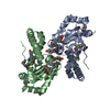


 PDBj
PDBj


 Assembly


 Mass: 54.938 Da / Num. of mol.: 2 / Source method: obtained synthetically / Formula: Mn
 Mass: 18.015 Da / Num. of mol.: 449 / Source method: isolated from a natural source / Formula: H2O
 Sample preparation
 / Beamline: 8.3.1 / Wavelength: 1.11
  Processing