[English] 日本語
Yorodumi
- PDB-1xan: HUMAN GLUTATHIONE REDUCTASE IN COMPLEX WITH A XANTHENE INHIBITOR -

+
Open data


ID or keywords:

Loading...

-
Basic information

Entry
Database: PDB / ID: 1xan
TitleHUMAN GLUTATHIONE REDUCTASE IN COMPLEX WITH A XANTHENE INHIBITOR
ComponentsGLUTATHIONE REDUCTASE
KeywordsGLUTATHIONE REDUCTASE / OXIDOREDUCTASE / FLAVOENZYME / GLUTATHIONE REDUCATASE
Function / homology
Function and homology information


glutathione-disulfide reductase / Metabolism of ingested H2SeO4 and H2SeO3 into H2Se / glutathione-disulfide reductase (NADPH) activity / Interconversion of nucleotide di- and triphosphates / NFE2L2 regulating anti-oxidant/detoxification enzymes / Detoxification of Reactive Oxygen Species / glutathione metabolic process / cell redox homeostasis / TP53 Regulates Metabolic Genes / NADP binding ...glutathione-disulfide reductase / Metabolism of ingested H2SeO4 and H2SeO3 into H2Se / glutathione-disulfide reductase (NADPH) activity / Interconversion of nucleotide di- and triphosphates / NFE2L2 regulating anti-oxidant/detoxification enzymes / Detoxification of Reactive Oxygen Species / glutathione metabolic process / cell redox homeostasis / TP53 Regulates Metabolic Genes / NADP binding / cellular response to oxidative stress / flavin adenine dinucleotide binding / electron transfer activity / mitochondrial matrix / external side of plasma membrane / mitochondrion / extracellular exosome / cytosol
Similarity search - Function
Glutathione reductase, eukaryote/bacterial / Pyridine nucleotide-disulphide oxidoreductase / : / Pyridine nucleotide-disulphide oxidoreductase, class I / FAD/NAD-linked reductase, C-terminal dimerisation domain / Pyridine nucleotide-disulphide oxidoreductase, class I, active site / Pyridine nucleotide-disulphide oxidoreductases class-I active site. / Pyridine nucleotide-disulphide oxidoreductase, dimerisation domain / Pyridine nucleotide-disulphide oxidoreductase, dimerisation domain / FAD/NAD-linked reductase, dimerisation domain superfamily ...Glutathione reductase, eukaryote/bacterial / Pyridine nucleotide-disulphide oxidoreductase / : / Pyridine nucleotide-disulphide oxidoreductase, class I / FAD/NAD-linked reductase, C-terminal dimerisation domain / Pyridine nucleotide-disulphide oxidoreductase, class I, active site / Pyridine nucleotide-disulphide oxidoreductases class-I active site. / Pyridine nucleotide-disulphide oxidoreductase, dimerisation domain / Pyridine nucleotide-disulphide oxidoreductase, dimerisation domain / FAD/NAD-linked reductase, dimerisation domain superfamily / FAD/NAD(P)-binding domain / Pyridine nucleotide-disulphide oxidoreductase / Enolase-like; domain 1 / FAD/NAD(P)-binding domain / FAD/NAD(P)-binding domain / 3-Layer(bba) Sandwich / FAD/NAD(P)-binding domain superfamily / 2-Layer Sandwich / Alpha Beta
Similarity search - Domain/homology
FLAVIN-ADENINE DINUCLEOTIDE / 3,6-DIHYDROXY-XANTHENE-9-PROPIONIC ACID / Glutathione reductase, mitochondrial
Similarity search - Component
Biological speciesHomo sapiens (human)
MethodX-RAY DIFFRACTION / Resolution: 2 Å
AuthorsSavvides, S.N. / Karplus, P.A.
CitationJournal: J.Biol.Chem. / Year: 1996
Title: Kinetics and crystallographic analysis of human glutathione reductase in complex with a xanthene inhibitor.
Authors: Savvides, S.N. / Karplus, P.A.
History
DepositionJan 26, 1996Processing site: BNL
Revision 1.0Jul 11, 1996Provider: repository / Type: Initial release
Revision 1.1Mar 24, 2008Group: Version format compliance
Revision 1.2Jul 13, 2011Group: Derived calculations / Version format compliance
Revision 1.3Jun 5, 2024Group: Data collection / Database references ...Data collection / Database references / Derived calculations / Other
Category: chem_comp_atom / chem_comp_bond ...chem_comp_atom / chem_comp_bond / database_2 / pdbx_database_status / pdbx_struct_special_symmetry / struct_site
Item: _database_2.pdbx_DOI / _database_2.pdbx_database_accession ..._database_2.pdbx_DOI / _database_2.pdbx_database_accession / _pdbx_database_status.process_site / _struct_site.pdbx_auth_asym_id / _struct_site.pdbx_auth_comp_id / _struct_site.pdbx_auth_seq_id

-
Structure visualization

Structure viewerMolecule:
MolmilJmol/JSmol

Downloads & links

-
Assembly

Deposited unit
A: GLUTATHIONE REDUCTASE
hetero molecules


Theoretical massNumber of molelcules
Total (without water)51,0503
Polymers49,9781
Non-polymers1,0722
Water9,314517
1
A: GLUTATHIONE REDUCTASE
hetero molecules

A: GLUTATHIONE REDUCTASE
hetero molecules


Theoretical massNumber of molelcules
Total (without water)102,1016
Polymers99,9572
Non-polymers2,1444
Water362
TypeNameSymmetry operationNumber
identity operation1_555x,y,z1
crystal symmetry operation2_656-x+1,y,-z+11
Buried area10860 Å2
ΔGint-54 kcal/mol
Surface area36940 Å2
MethodPISA, PQS
Unit cell
Length a, b, c (Å)119.790, 63.350, 84.650
Angle α, β, γ (deg.)90.00, 58.61, 90.00
Int Tables number5
Space group name H-MC121
Components on special symmetry positions
IDModelComponents
11A-1024-

HXP

21A-1024-

HXP

31A-1024-

HXP

-
Components

#1: Protein GLUTATHIONE REDUCTASE /


Mass: 49978.430 Da / Num. of mol.: 1
Source method: isolated from a genetically manipulated source
Source: (gene. exp.) Homo sapiens (human) / Production host: Escherichia coli (E. coli) / Strain (production host): SG5 / References: UniProt: P00390, EC: 1.6.4.2
#2: Chemical ChemComp-FAD / FLAVIN-ADENINE DINUCLEOTIDE / Flavin adenine dinucleotide


Mass: 785.550 Da / Num. of mol.: 1 / Source method: obtained synthetically / Formula: C27H33N9O15P2 / Comment: FAD*YM
#3: Chemical ChemComp-HXP / 3,6-DIHYDROXY-XANTHENE-9-PROPIONIC ACID


Mass: 286.279 Da / Num. of mol.: 1 / Source method: obtained synthetically / Formula: C16H14O5
#4: Water ChemComp-HOH / water / Water


Mass: 18.015 Da / Num. of mol.: 517 / Source method: isolated from a natural source / Formula: H2O
Compound details
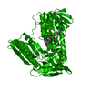
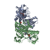
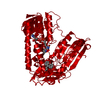
IN THE INHIBITOR-FREE CRYSTAL STRUCTURE OF HUMAN GLUTATHIONE REDUCTASE (PDB ENTRY 3GRS) AN ...IN THE INHIBITOR-FREE CRYSTAL STRUCTURE OF HUMAN GLUTATHIONE REDUCTASE (PDB ENTRY 3GRS) AN INTERSUBUNIT DISULFIDE BOND OF OPTIMAL GEOMETRY IS OBSERVED BETWEEN CYS 90 AND ITS CRYSTALLOGRAPHIC TWO-FOLD RELATED MATE. IN THIS ENTRY, HOWEVER, THIS INTERACTION IS NOT AS OPTIMAL DUE TO THE PERTURBATIONS IN THE PROTEIN STRUCTURE RESULTING FROM INHIBITOR BINDING. THE ELECTRON DENSITY CORRESPONDING TO THE REGION OF THE DISULFIDE BRIDGE IN QUESTION IS LESS AND ELONGATED WHEN COMPARED WITH THAT OF THE INHIBITOR-FREE STRUCTURE. THIS AGRESS WITH THE INCREASED MOBILITY OF THIS REGION IN THE INHIBITOR-BOUND STRUCTURE AND SUGGESTS A POSSIBLE DISRUPTION OF THE INTERSUBUNIT DISULFIDE BRIDGE IN FRACTION OF THE MOLECULES. HYDROGEN BONDING INTERACTIONS IN THIS REGION FURTHER SUPPORT A PARTIALLY OPEN CONFORMATION FOR THE CYS 90 - CYS90' DISULFIDE BOND. FOR A MORE EXTENSIVE DISCUSSION PLEASE CONSULT THE JRNL REFERENCE.

-
Experimental details

-
Experiment

ExperimentMethod: X-RAY DIFFRACTION

-
Sample preparation

CrystalDensity Matthews: 2.74 Å3/Da / Density % sol: 55.14 %
Crystal grow
*PLUS
pH: 7 / Method: vapor diffusion, hanging drop
Components of the solutions
*PLUS
IDConc.Common nameCrystal-IDSol-ID
120 mg/mlhGR1drop
23 %ammonium sulfate1drop
30.1 Mpotassium phosphate1drop
40.1 %beta-octylglucoside1drop
521-23 %ammonium sulfate1reservoir
60.1 Mpotassium phosphate1reservoir

-
Data collection

RadiationScattering type: x-ray
Radiation wavelengthRelative weight: 1
Reflection
*PLUS
Highest resolution: 2 Å / Num. obs: 33736 / % possible obs: 92 % / Rmerge(I) obs: 0.071

-
Processing

Software
NameClassification
X-PLORmodel building
X-PLORrefinement
X-PLORphasing
RefinementResolution: 2→10 Å / σ(F): 0
RfactorNum. reflection% reflection
Rwork0.158 --
obs0.158 33736 92 %
Displacement parametersBiso mean: 2 Å2
Refinement stepCycle: LAST / Resolution: 2→10 Å
ProteinNucleic acidLigandSolventTotal
Num. atoms3499 0 74 517 4090

+
About Yorodumi

-
News

-
Feb 9, 2022. New format data for meta-information of EMDB entries

New format data for meta-information of EMDB entries

  • Version 3 of the EMDB header file is now the official format.
  • The previous official version 1.9 will be removed from the archive.

Related info.:EMDB header

External links:wwPDB to switch to version 3 of the EMDB data model

-
Aug 12, 2020. Covid-19 info

Covid-19 info

URL: https://pdbj.org/emnavi/covid19.php

New page: Covid-19 featured information page in EM Navigator.

Related info.:Covid-19 info / Mar 5, 2020. Novel coronavirus structure data

+
Mar 5, 2020. Novel coronavirus structure data

Novel coronavirus structure data

Related info.:Yorodumi Speices / Aug 12, 2020. Covid-19 info

External links:COVID-19 featured content - PDBj / Molecule of the Month (242):Coronavirus Proteases

+
Jan 31, 2019. EMDB accession codes are about to change! (news from PDBe EMDB page)

EMDB accession codes are about to change! (news from PDBe EMDB page)

  • The allocation of 4 digits for EMDB accession codes will soon come to an end. Whilst these codes will remain in use, new EMDB accession codes will include an additional digit and will expand incrementally as the available range of codes is exhausted. The current 4-digit format prefixed with “EMD-” (i.e. EMD-XXXX) will advance to a 5-digit format (i.e. EMD-XXXXX), and so on. It is currently estimated that the 4-digit codes will be depleted around Spring 2019, at which point the 5-digit format will come into force.
  • The EM Navigator/Yorodumi systems omit the EMD- prefix.

Related info.:Q: What is EMD? / ID/Accession-code notation in Yorodumi/EM Navigator

External links:EMDB Accession Codes are Changing Soon! / Contact to PDBj

+
Jul 12, 2017. Major update of PDB

Major update of PDB

  • wwPDB released updated PDB data conforming to the new PDBx/mmCIF dictionary.
  • This is a major update changing the version number from 4 to 5, and with Remediation, in which all the entries are updated.
  • In this update, many items about electron microscopy experimental information are reorganized (e.g. em_software).
  • Now, EM Navigator and Yorodumi are based on the updated data.

External links:wwPDB Remediation / Enriched Model Files Conforming to OneDep Data Standards Now Available in the PDB FTP Archive

-
Yorodumi

Thousand views of thousand structures

  • Yorodumi is a browser for structure data from EMDB, PDB, SASBDB, etc.
  • This page is also the successor to EM Navigator detail page, and also detail information page/front-end page for Omokage search.
  • The word "yorodu" (or yorozu) is an old Japanese word meaning "ten thousand". "mi" (miru) is to see.

Related info.:EMDB / PDB / SASBDB / Comparison of 3 databanks / Yorodumi Search / Aug 31, 2016. New EM Navigator & Yorodumi / Yorodumi Papers / Jmol/JSmol / Function and homology information / Changes in new EM Navigator and Yorodumi

Read more