 Movie
Movie Controller

[English] 日本語
 Yorodumi
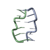
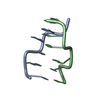
- PDB-1xam: Cobalt hexammine induced tautameric shift in Z-DNA: structure of ... -
+ Open data

- Basic information
| Entry | Database: PDB / ID: 1xam | ||||||
|---|---|---|---|---|---|---|---|
| Title | Cobalt hexammine induced tautameric shift in Z-DNA: structure of d(CGCGCA).d(TGCGCG) in two crystal forms. | ||||||
 Components Components |
| ||||||
 Keywords Keywords |  DNA / DOUBLE HELIX / Z-DNA DNA / DOUBLE HELIX / Z-DNA | ||||||
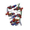
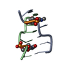
| Function / homology | COBALT HEXAMMINE(III) / DNA Function and homology information Function and homology information | ||||||
| Method | X-RAY DIFFRACTION / MOLECULAR REPLACEMENT / Resolution: 1.86 Å | ||||||
 Authors Authors | Thiyagarajan, S. / Rajan, S.S. / Gautham, N. | ||||||
 Citation Citation | Journal: Nucleic Acids Res. / Year: 2004 Title: Cobalt hexammine induced tautomeric shift in Z-DNA: the structure of d(CGCGCA)*d(TGCGCG) in two crystal forms. Authors: Thiyagarajan, S. / Rajan, S.S. / Gautham, N. #1: Journal: Acta Crystallogr.,Sect.D / Year: 2005 Title: Structure of d(TGCGCG). d(CGCGCA) in two crystal forms: effects of sequence and crystal packing in Z-DNA Authors: Thiyagarajan, S. / Rajan, S.S. / Gautham, N. | ||||||
| History |
|
- Structure visualization
| Structure viewer | Molecule: MolmilJmol/JSmol |
|---|
- Downloads & links
-Download
| PDBx/mmCIF format | 1xam.cif.gz | 19 KB | Display | PDBx/mmCIF format |
|---|---|---|---|---|
| PDB format | pdb1xam.ent.gz | 12.3 KB | Display | PDB format |
| PDBx/mmJSON format | 1xam.json.gz | Tree view | PDBx/mmJSON format | |
| Others |  Other downloads Other downloads |
-Validation report
| Arichive directory | https://data.pdbj.org/pub/pdb/validation_reports/xa/1xamftp://data.pdbj.org/pub/pdb/validation_reports/xa/1xam | HTTPS FTP |
|---|
-Related structure data


-Links
 PDBj
PDBj


- Assembly
| Deposited unit | 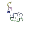
| ||||||||
|---|---|---|---|---|---|---|---|---|---|
| 1 | 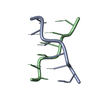
| ||||||||
| 2 | 
| ||||||||
| Unit cell |
| ||||||||
| Details | biological unit is a double helix. A second hexamer is to be generated by symmetry related entities of the dinucleotide formed with chain C and D. |
-Components
| #1: DNA chain | Mass: 1794.206 Da / Num. of mol.: 1 / Source method: obtained synthetically | ||||
|---|---|---|---|---|---|
| #2: DNA chain | Mass: 1825.216 Da / Num. of mol.: 1 / Source method: obtained synthetically | ||||
| #3: DNA chain | Mass: 588.441 Da / Num. of mol.: 2 / Source method: obtained synthetically #4: Chemical | ChemComp-NCO / |   Mass: 161.116 Da / Num. of mol.: 1 / Source method: obtained synthetically / Formula: CoH18N6 Mass: 161.116 Da / Num. of mol.: 1 / Source method: obtained synthetically / Formula: CoH18N6#5: Water | ChemComp-HOH / | Water Mass: 18.015 Da / Num. of mol.: 18 / Source method: isolated from a natural source / Formula: H2O Mass: 18.015 Da / Num. of mol.: 18 / Source method: isolated from a natural source / Formula: H2O |
-Experimental details
-Experiment
| Experiment | Method: X-RAY DIFFRACTION / Number of used crystals: 1 |
|---|
- Sample preparation
| Crystal | Density Matthews: 1.76 Å3/Da / Density % sol: 29.41 % | ||||||||||||||||||||||||||||||||||||
|---|---|---|---|---|---|---|---|---|---|---|---|---|---|---|---|---|---|---|---|---|---|---|---|---|---|---|---|---|---|---|---|---|---|---|---|---|---|
| Crystal grow | Temperature: 293 K / Method: vapor diffusion, hanging drop / pH: 6.9 Details: MPD, cacodylate, cobalt hexammine chloride, spermine., pH 6.9, VAPOR DIFFUSION, HANGING DROP, temperature 293K | ||||||||||||||||||||||||||||||||||||
| Components of the solutions |
|
-Data collection
| Diffraction | Mean temperature: 293 K |
|---|---|
| Diffraction source | Source: ROTATING ANODE / Type: RIGAKU RU300 / Wavelength: 1.5418 Å |
| Detector | Type: MARRESEARCH / Detector: IMAGE PLATE / Date: Jan 25, 2004 |
| Radiation | Monochromator: NIL / Protocol: SINGLE WAVELENGTH / Monochromatic (M) / Laue (L): M / Scattering type: x-ray |
| Radiation wavelength | Wavelength: 1.5418 Å / Relative weight: 1 |
| Reflection | Resolution: 1.86→30 Å / Num. all: 2695 / Num. obs: 2686 / % possible obs: 99 % / Observed criterion σ(F): 0 / Observed criterion σ(I): 0 / Redundancy: 4.64 % / Rmerge(I) obs: 0.15 / Net I/σ(I): 2.4 |
| Reflection shell | Resolution: 1.86→1.93 Å / Redundancy: 4.34 % / Rmerge(I) obs: 0.5274 / Mean I/σ(I) obs: 1 / % possible all: 90.3 |
- Processing
| Software |
| |||||||||||||||||||||||||||||||||||||||||||||||||||||||
|---|---|---|---|---|---|---|---|---|---|---|---|---|---|---|---|---|---|---|---|---|---|---|---|---|---|---|---|---|---|---|---|---|---|---|---|---|---|---|---|---|---|---|---|---|---|---|---|---|---|---|---|---|---|---|---|---|
| Refinement | Method to determine structure: MOLECULAR REPLACEMENT Starting model: Z-DNA hexamer with terminal base pairs replaced with A-T base pair. Resolution: 1.86→30 Å / Cor.coef. Fo:Fc: 0.935 / Cor.coef. Fo:Fc free: 0.879 / SU B: 7.847 / SU ML: 0.219 / Cross valid method: THROUGHOUT / σ(F): 0 / σ(I): 0 / ESU R: 0.294 / ESU R Free: 0.248 / Stereochemistry target values: MAXIMUM LIKELIHOOD
| |||||||||||||||||||||||||||||||||||||||||||||||||||||||
| Solvent computation | Ion probe radii: 0.8 Å / Shrinkage radii: 0.8 Å / VDW probe radii: 1.4 Å / Solvent model: BABINET MODEL WITH MASK | |||||||||||||||||||||||||||||||||||||||||||||||||||||||
| Displacement parameters | Biso mean: 24.41 Å2
| |||||||||||||||||||||||||||||||||||||||||||||||||||||||
| Refinement step | Cycle: LAST / Resolution: 1.86→30 Å
| |||||||||||||||||||||||||||||||||||||||||||||||||||||||
| Refine LS restraints |
| |||||||||||||||||||||||||||||||||||||||||||||||||||||||
| LS refinement shell | Resolution: 1.86→1.909 Å / Total num. of bins used: 20 /
|
