 Movie
Movie Controller

[English] 日本語
 Yorodumi
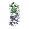
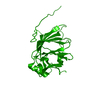
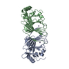
- PDB-1x8e: Crystal structure of Pyrococcus furiosus phosphoglucose isomerase... -
+ Open data

- Basic information
| Entry | Database: PDB / ID: 1x8e | ||||||
|---|---|---|---|---|---|---|---|
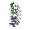
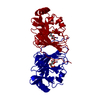
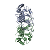
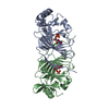
| Title | Crystal structure of Pyrococcus furiosus phosphoglucose isomerase free enzyme | ||||||
 Components Components | Glucose-6-phosphate isomerase | ||||||
 Keywords Keywords | ISOMERASE / CUPIN SUPERFAMILY / PYROCOCCUS FURIOSUS / HYPERTHERMOPHILE / PHOSPHOGLUCOSE ISOMERASE / EXTREMEOPHILE | ||||||
| Function / homology |  Function and homology informationglucose-6-phosphate isomerase / glucose-6-phosphate isomerase activity / gluconeogenesis / glycolytic process / iron ion binding / cytoplasm Function and homology informationglucose-6-phosphate isomerase / glucose-6-phosphate isomerase activity / gluconeogenesis / glycolytic process / iron ion binding / cytoplasmSimilarity search - Function | ||||||
| Biological species |   Pyrococcus furiosus (archaea) Pyrococcus furiosus (archaea) | ||||||
| Method | X-RAY DIFFRACTION / MOLECULAR REPLACEMENT / Resolution: 2.8 Å | ||||||
 Authors Authors | Berrisford, J.M. / Akerboom, J. / Brouns, S. / Sedelnikova, S.E. / Turnbull, A.P. / van der Oost, J. / Salmon, L. / Hardre, R. / Murray, I.A. / Blackburn, G.M. ...Berrisford, J.M. / Akerboom, J. / Brouns, S. / Sedelnikova, S.E. / Turnbull, A.P. / van der Oost, J. / Salmon, L. / Hardre, R. / Murray, I.A. / Blackburn, G.M. / Rice, D.W. / Baker, P.J. | ||||||
 Citation Citation | Journal: J.Mol.Biol. / Year: 2004 Title: The structures of inhibitor complexes of Pyrococcus furiosus phosphoglucose isomerase provide insights into substrate binding and catalysis. Authors: Berrisford, J.M. / Akerboom, J. / Brouns, S. / Sedelnikova, S.E. / Turnbull, A.P. / van der Oost, J. / Salmon, L. / Hardre, R. / Murray, I.A. / Blackburn, G.M. / Rice, D.W. / Baker, P.J. | ||||||
| History |
|
- Structure visualization
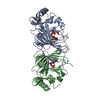
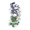
| Structure viewer | Molecule: MolmilJmol/JSmol |
|---|
- Downloads & links
-Download
| PDBx/mmCIF format | 1x8e.cif.gz | 85 KB | Display | PDBx/mmCIF format |
|---|---|---|---|---|
| PDB format | pdb1x8e.ent.gz | 65.7 KB | Display | PDB format |
| PDBx/mmJSON format | 1x8e.json.gz | Tree view | PDBx/mmJSON format | |
| Others |  Other downloads Other downloads |
-Validation report
| Arichive directory | https://data.pdbj.org/pub/pdb/validation_reports/x8/1x8eftp://data.pdbj.org/pub/pdb/validation_reports/x8/1x8e | HTTPS FTP |
|---|
-Related structure data
| Related structure data |  1x7nC 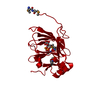 1x82SC S: Starting model for refinement C: citing same article ( |
|---|---|
| Similar structure data |
-Links
 PDBj
PDBj

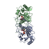
- Assembly
| Deposited unit | 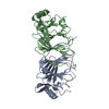
| ||||||||
|---|---|---|---|---|---|---|---|---|---|
| 1 |
| ||||||||
| Unit cell |
| ||||||||
| Details | Subunits A and B generate the biological dimer |
-Components
| #1: Protein | / GPI / Phosphoglucose isomerase / PGI / Phosphohexose isomerase / PHI Mass: 21636.633 Da / Num. of mol.: 2 Source method: isolated from a genetically manipulated source Source: (gene. exp.) Pyrococcus furiosus (archaea) / Gene: pgiA / Plasmid: PET24-d / Species (production host): Escherichia coli / Production host:  Escherichia coli BL21(DE3) (bacteria) / Strain (production host): BL21(DE3) / References: UniProt: P83194, glucose-6-phosphate isomerase Escherichia coli BL21(DE3) (bacteria) / Strain (production host): BL21(DE3) / References: UniProt: P83194, glucose-6-phosphate isomerase |
|---|
-Experimental details
-Experiment
| Experiment | Method: X-RAY DIFFRACTION / Number of used crystals: 1 |
|---|
- Sample preparation
| Crystal | Density Matthews: 2.1 Å3/Da / Density % sol: 41.7 % |
|---|---|
| Crystal grow | Temperature: 290 K / Method: vapor diffusion, hanging drop / pH: 6.5 Details: 1.6M tri-sodium citrate, pH 6.5, VAPOR DIFFUSION, HANGING DROP, temperature 290K |
-Data collection
| Diffraction | Mean temperature: 100 K |
|---|---|
| Diffraction source | Source: ROTATING ANODE / Type: RIGAKU RU200 / Wavelength: 1.54 Å |
| Detector | Type: MARRESEARCH / Detector: IMAGE PLATE / Date: Aug 27, 2003 / Details: YALE MIRRORS |
| Radiation | Protocol: SINGLE WAVELENGTH / Monochromatic (M) / Laue (L): M / Scattering type: x-ray |
| Radiation wavelength | Wavelength: 1.54 Å / Relative weight: 1 |
| Reflection | Resolution: 2.8→53.45 Å / Num. all: 10198 / Num. obs: 10096 / % possible obs: 99 % / Redundancy: 4.1 % / Rmerge(I) obs: 0.141 / Net I/σ(I): 4.9 |
| Reflection shell | Resolution: 2.8→2.95 Å / Redundancy: 4.1 % / Rmerge(I) obs: 0.439 / Mean I/σ(I) obs: 1.7 / Num. unique all: 1359 / % possible all: 4.1 |
- Processing
| Software |
| ||||||||||||||||||||||||||||||||||||||||||||||||||||||||||||||||||||||||||||||||||||||||||||||||||||
|---|---|---|---|---|---|---|---|---|---|---|---|---|---|---|---|---|---|---|---|---|---|---|---|---|---|---|---|---|---|---|---|---|---|---|---|---|---|---|---|---|---|---|---|---|---|---|---|---|---|---|---|---|---|---|---|---|---|---|---|---|---|---|---|---|---|---|---|---|---|---|---|---|---|---|---|---|---|---|---|---|---|---|---|---|---|---|---|---|---|---|---|---|---|---|---|---|---|---|---|---|---|
| Refinement | Method to determine structure: MOLECULAR REPLACEMENT Starting model: 1X82 Resolution: 2.8→53.45 Å / Cor.coef. Fo:Fc: 0.913 / Cor.coef. Fo:Fc free: 0.85 / SU B: 19.835 / SU ML: 0.373 / Cross valid method: THROUGHOUT / ESU R Free: 0.447 / Stereochemistry target values: MAXIMUM LIKELIHOOD / Details: HYDROGENS HAVE BEEN ADDED IN THE RIDING POSITIONS
| ||||||||||||||||||||||||||||||||||||||||||||||||||||||||||||||||||||||||||||||||||||||||||||||||||||
| Solvent computation | Ion probe radii: 0.8 Å / Shrinkage radii: 0.8 Å / VDW probe radii: 1.4 Å / Solvent model: BABINET MODEL WITH MASK | ||||||||||||||||||||||||||||||||||||||||||||||||||||||||||||||||||||||||||||||||||||||||||||||||||||
| Displacement parameters | Biso mean: 24.614 Å2
| ||||||||||||||||||||||||||||||||||||||||||||||||||||||||||||||||||||||||||||||||||||||||||||||||||||
| Refinement step | Cycle: LAST / Resolution: 2.8→53.45 Å
| ||||||||||||||||||||||||||||||||||||||||||||||||||||||||||||||||||||||||||||||||||||||||||||||||||||
| Refine LS restraints |
| ||||||||||||||||||||||||||||||||||||||||||||||||||||||||||||||||||||||||||||||||||||||||||||||||||||
| LS refinement shell | Resolution: 2.8→2.873 Å / Total num. of bins used: 20
|
