 Movie
Movie Controller

[English] 日本語
 Yorodumi
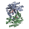
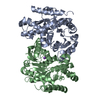
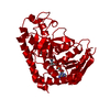
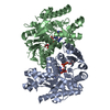
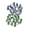
- PDB-1wzi: Structural basis for alteration of cofactor specificity of Malate... -
+ Open data

- Basic information
| Entry | Database: PDB / ID: 1wzi | ||||||
|---|---|---|---|---|---|---|---|
| Title | Structural basis for alteration of cofactor specificity of Malate dehydrogenase from Thermus flavus | ||||||
 Components Components | Malate dehydrogenase | ||||||
 Keywords Keywords | OXIDOREDUCTASE / Seven amino acid residues mutant / protein-NADPH complex | ||||||
| Function / homology |  Function and homology informationmalate dehydrogenase / L-malate dehydrogenase activity / malate metabolic process / tricarboxylic acid cycle Function and homology informationmalate dehydrogenase / L-malate dehydrogenase activity / malate metabolic process / tricarboxylic acid cycleSimilarity search - Function | ||||||
| Biological species |   Thermus thermophilus (bacteria) Thermus thermophilus (bacteria) | ||||||
| Method | X-RAY DIFFRACTION / SYNCHROTRON / MOLECULAR REPLACEMENT / Resolution: 2 Å | ||||||
 Authors Authors | Tomita, T. / Fushinobu, S. / Kuzuyama, T. / Nishiyama, M. | ||||||
 Citation Citation | Journal: to be published Title: Structural basis for alteration of cofactor specificity of malate dehydrogenase from Thermus flavus Authors: Tomita, T. / Fushinobu, S. / Kuzuyama, T. / Nishiyama, M. #1: Journal: Biochemistry / Year: 1993Title: Determinants of protein thermostability observed in the 1.9 Angstroms crystal structure of malate dehydrogenase from the thermophilic bacterium Thermus flavus Authors: Kelly, C.A. / Nishiyama, M. / Ohnishi, Y. / Beppu, T. / Birktoft, J.J. #2: Journal: J.Mol.Biol. / Year: 1991 Title: Preliminary X-ray diffraction analysis of a crystallizable mutant of malate dehydrogenase from the thermophile Thermus flavus Authors: Kelly, C.A. / Sarfaty, S. / Nishiyama, M. / Beppu, T. / Birktoft, J.J. #3: Journal: Biochemistry / Year: 1989Title: Refined crystal structure of cytoplasmic malate dehydrogenase at 2.5-A resolution Authors: Birktoft, J.J. / Rhodes, G. / Banaszak, L.J. #4: Journal: J.Biol.Chem. / Year: 1986 Title: Nucleotide sequence of the malate dehydrogenase gene of Thermus flavus and its mutation directing an increase in enzyme activity Authors: Nishiyama, M. / Matsubara, N. / Yamamoto, K. / Iijima, S. / Uozumi, T. / Beppu, T. #5: Journal: J.Biol.Chem. / Year: 1983 Title: The presence of a histidine-aspartic acid pair in the active site of 2-hydroxyacid dehydrogenases. X-ray refinement of cytoplasmic malate dehydrogenase Authors: Birktoft, J.J. / Banaszak, L.J. #6: Journal: Enzyme / Year: 1975Title: Malate Dehydrogenases Authors: Banaszak, L.J. / Bradshaw, R.A. | ||||||
| History |
|
- Structure visualization
| Structure viewer | Molecule: MolmilJmol/JSmol |
|---|
- Downloads & links
-Download
| PDBx/mmCIF format | 1wzi.cif.gz | 152.3 KB | Display | PDBx/mmCIF format |
|---|---|---|---|---|
| PDB format | pdb1wzi.ent.gz | 117.7 KB | Display | PDB format |
| PDBx/mmJSON format | 1wzi.json.gz | Tree view | PDBx/mmJSON format | |
| Others |  Other downloads Other downloads |
-Validation report
| Arichive directory | https://data.pdbj.org/pub/pdb/validation_reports/wz/1wziftp://data.pdbj.org/pub/pdb/validation_reports/wz/1wzi | HTTPS FTP |
|---|
-Related structure data
| Related structure data |  1wzeC 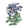 1bmdS S: Starting model for refinement C: citing same article ( |
|---|---|
| Similar structure data |


-Links
 PDBj
PDBj


- Assembly
| Deposited unit | 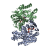
| ||||||||
|---|---|---|---|---|---|---|---|---|---|
| 1 |
| ||||||||
| Unit cell |
| ||||||||
| Details | The second part of the biological assembly is generated by the two fold axis: -x+1, y, -z. |
-Components
| #1: Protein | Mass: 35460.637 Da / Num. of mol.: 2 / Mutation: E41G, I42S, P43E, Q44R, A45S, M46F, K47Q Source method: isolated from a genetically manipulated source Details: Malate dehydrogenase mutant EX7 / Source: (gene. exp.) Thermus thermophilus (bacteria) / Strain: AT-62 / Gene: mdh / Plasmid: pUC19 / Production host: Escherichia coli (E. coli) / Strain (production host): JM109 / References: UniProt: P10584, malate dehydrogenase#2: Chemical | Nicotinamide adenine dinucleotide phosphate  Mass: 745.421 Da / Num. of mol.: 2 / Source method: obtained synthetically / Formula: C21H30N7O17P3 Mass: 745.421 Da / Num. of mol.: 2 / Source method: obtained synthetically / Formula: C21H30N7O17P3#3: Water | ChemComp-HOH / | Water Mass: 18.015 Da / Num. of mol.: 624 / Source method: isolated from a natural source / Formula: H2O Mass: 18.015 Da / Num. of mol.: 624 / Source method: isolated from a natural source / Formula: H2O |
|---|
-Experimental details
-Experiment
| Experiment | Method: X-RAY DIFFRACTION / Number of used crystals: 1 |
|---|
- Sample preparation
| Crystal | Density Matthews: 2.5 Å3/Da / Density % sol: 50.33 % |
|---|---|
| Crystal grow | Temperature: 293 K / Method: vapor diffusion, hanging drop / pH: 8.5 Details: PEG 4000, dithiothreitol, pH 8.5, VAPOR DIFFUSION, HANGING DROP, temperature 293K |
-Data collection
| Diffraction | Mean temperature: 95 K |
|---|---|
| Diffraction source | Source: SYNCHROTRON / Site: Photon Factory  / Beamline: BL-6A / Wavelength: 1 Å / Beamline: BL-6A / Wavelength: 1 Å |
| Detector | Type: ADSC QUANTUM 4 / Detector: CCD / Date: Oct 24, 2003 |
| Radiation | Protocol: SINGLE WAVELENGTH / Monochromatic (M) / Laue (L): M / Scattering type: x-ray |
| Radiation wavelength | Wavelength: 1 Å / Relative weight: 1 |
| Reflection | Resolution: 2→28.89 Å / Num. all: 48624 / Num. obs: 48624 / % possible obs: 99.7 % / Observed criterion σ(F): 2 / Observed criterion σ(I): 2 / Biso Wilson estimate: 8.6 Å2 / Rmerge(I) obs: 0.05 / Rsym value: 0.05 |
| Reflection shell | Resolution: 2→2.13 Å / % possible all: 100 |
- Processing
| Software |
| ||||||||||||||||||||||||||||||||||||
|---|---|---|---|---|---|---|---|---|---|---|---|---|---|---|---|---|---|---|---|---|---|---|---|---|---|---|---|---|---|---|---|---|---|---|---|---|---|
| Refinement | Method to determine structure: MOLECULAR REPLACEMENT Starting model: PDB ENTRY 1BMD Resolution: 2→28.89 Å / Rfactor Rfree error: 0.005 / Data cutoff high absF: 2286096.97 / Data cutoff low absF: 0 / Isotropic thermal model: RESTRAINED / Cross valid method: THROUGHOUT / σ(F): 0 / Stereochemistry target values: Engh & Huber
| ||||||||||||||||||||||||||||||||||||
| Solvent computation | Solvent model: FLAT MODEL / Bsol: 45.7329 Å2 / ksol: 0.350692 e/Å3 | ||||||||||||||||||||||||||||||||||||
| Displacement parameters | Biso mean: 18.7 Å2
| ||||||||||||||||||||||||||||||||||||
| Refine analyze |
| ||||||||||||||||||||||||||||||||||||
| Refinement step | Cycle: LAST / Resolution: 2→28.89 Å
| ||||||||||||||||||||||||||||||||||||
| Refine LS restraints |
| ||||||||||||||||||||||||||||||||||||
| LS refinement shell | Resolution: 2→2.13 Å / Rfactor Rfree error: 0.012 / Total num. of bins used: 6
| ||||||||||||||||||||||||||||||||||||
| Xplor file |
|
