 Movie
Movie Controller

[English] 日本語
 Yorodumi
- PDB-1wxq: Crystal Structure of GTP binding protein from Pyrococcus horikosh... -
+ Open data

- Basic information
| Entry | Database: PDB / ID: 1wxq | ||||||
|---|---|---|---|---|---|---|---|
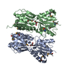
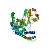
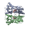
| Title | Crystal Structure of GTP binding protein from Pyrococcus horikoshii OT3 | ||||||
 Components Components | GTP-binding protein G protein G protein | ||||||
 Keywords Keywords | STRUCTURAL GENOMICS / UNKNOWN FUNCTION / GTP-binding protein / RIKEN Structural Genomics/Proteomics Initiative / RSGI / NPPSFA / National Project on Protein Structural and Functional Analyses | ||||||
| Function / homology |  Function and homology information Function and homology information | ||||||
| Biological species |   Pyrococcus horikoshii (archaea) Pyrococcus horikoshii (archaea) | ||||||
| Method | X-RAY DIFFRACTION / SYNCHROTRON / MAD / Resolution: 2.6 Å | ||||||
 Authors Authors | Lokanath, N.K. / Kunishima, N. / RIKEN Structural Genomics/Proteomics Initiative (RSGI) | ||||||
 Citation Citation | Journal: To be Published Title: Crystal Structure of GTP binding protein from Pyrococcus horikoshii OT3 Authors: Lokanath, N.K. / Kunishima, N. | ||||||
| History |
|
- Structure visualization
| Structure viewer | Molecule: MolmilJmol/JSmol |
|---|
- Downloads & links
-Download
| PDBx/mmCIF format | 1wxq.cif.gz | 79.2 KB | Display | PDBx/mmCIF format |
|---|---|---|---|---|
| PDB format | pdb1wxq.ent.gz | 63.7 KB | Display | PDB format |
| PDBx/mmJSON format | 1wxq.json.gz | Tree view | PDBx/mmJSON format | |
| Others |  Other downloads Other downloads |
-Validation report
| Arichive directory | https://data.pdbj.org/pub/pdb/validation_reports/wx/1wxqftp://data.pdbj.org/pub/pdb/validation_reports/wx/1wxq | HTTPS FTP |
|---|
-Related structure data
| Similar structure data | |
|---|---|
| Other databases |


-Links
 PDBj
PDBj


- Assembly
| Deposited unit | 
| ||||||||
|---|---|---|---|---|---|---|---|---|---|
| 1 |
| ||||||||
| Unit cell |
| ||||||||
| Details | biological assembly is monomer |
-Components
| #1: Protein | G protein Mass: 44846.109 Da / Num. of mol.: 1 Source method: isolated from a genetically manipulated source Source: (gene. exp.) Pyrococcus horikoshii (archaea) / Strain: OT3 / Plasmid: pET11a / Production host:  Escherichia coli (E. coli) / Strain (production host): 21-CodonPlus (DE3)-RIL / References: UniProt: O58261 Escherichia coli (E. coli) / Strain (production host): 21-CodonPlus (DE3)-RIL / References: UniProt: O58261 |
|---|---|
| #2: Water | ChemComp-HOH / Water Mass: 18.015 Da / Num. of mol.: 89 / Source method: isolated from a natural source / Formula: H2O Mass: 18.015 Da / Num. of mol.: 89 / Source method: isolated from a natural source / Formula: H2O |
-Experimental details
-Experiment
| Experiment | Method: X-RAY DIFFRACTION / Number of used crystals: 1 |
|---|
- Sample preparation
| Crystal | Density Matthews: 2.4 Å3/Da / Density % sol: 47.9 % |
|---|---|
| Crystal grow | Temperature: 295 K / Method: microbatch / pH: 7.5 Details: PEG 4000, HEPES, Dioxane, pH 7.5, microbatch, temperature 295K |
-Data collection
| Diffraction | Mean temperature: 100 K | ||||||||||||
|---|---|---|---|---|---|---|---|---|---|---|---|---|---|
| Diffraction source | Source: SYNCHROTRON / Site: SPring-8  / Beamline: BL26B1 / Wavelength: 0.9791, 0.97942, 1.0 / Beamline: BL26B1 / Wavelength: 0.9791, 0.97942, 1.0 | ||||||||||||
| Detector | Type: RIGAKU RAXIS / Detector: IMAGE PLATE / Date: Nov 6, 2004 / Details: RH coated bent cylindrical mirror | ||||||||||||
| Radiation | Monochromator: Si111 double crystal / Protocol: MAD / Monochromatic (M) / Laue (L): M / Scattering type: x-ray | ||||||||||||
| Radiation wavelength |
| ||||||||||||
| Reflection | Resolution: 2.6→30 Å / Num. all: 11882 / Num. obs: 11523 / % possible obs: 99.1 % / Observed criterion σ(F): 0 / Observed criterion σ(I): 0 / Redundancy: 3 % / Biso Wilson estimate: 32.42 Å2 / Rmerge(I) obs: 0.076 / Net I/σ(I): 13.2 | ||||||||||||
| Reflection shell | Resolution: 2.6→2.69 Å / % possible all: 98.9 |
- Processing
| Software |
| ||||||||||||||||||||||||||||||||||||||||||||||||||||||||||||||||||||||
|---|---|---|---|---|---|---|---|---|---|---|---|---|---|---|---|---|---|---|---|---|---|---|---|---|---|---|---|---|---|---|---|---|---|---|---|---|---|---|---|---|---|---|---|---|---|---|---|---|---|---|---|---|---|---|---|---|---|---|---|---|---|---|---|---|---|---|---|---|---|---|---|
| Refinement | Method to determine structure: MAD / Resolution: 2.6→10 Å / Cor.coef. Fo:Fc: 0.879 / Cor.coef. Fo:Fc free: 0.845 / SU B: 14.61 / SU ML: 0.326 / TLS residual ADP flag: LIKELY RESIDUAL / Cross valid method: THROUGHOUT / σ(F): 0 / ESU R: 1.927 / ESU R Free: 0.403 / Stereochemistry target values: MAXIMUM LIKELIHOOD
| ||||||||||||||||||||||||||||||||||||||||||||||||||||||||||||||||||||||
| Solvent computation | Ion probe radii: 0.8 Å / Shrinkage radii: 0.8 Å / VDW probe radii: 1.4 Å / Solvent model: BABINET MODEL WITH MASK | ||||||||||||||||||||||||||||||||||||||||||||||||||||||||||||||||||||||
| Displacement parameters | Biso mean: 39.842 Å2
| ||||||||||||||||||||||||||||||||||||||||||||||||||||||||||||||||||||||
| Refinement step | Cycle: LAST / Resolution: 2.6→10 Å
| ||||||||||||||||||||||||||||||||||||||||||||||||||||||||||||||||||||||
| Refine LS restraints |
| ||||||||||||||||||||||||||||||||||||||||||||||||||||||||||||||||||||||
| LS refinement shell | Resolution: 2.6→2.663 Å / Total num. of bins used: 20 /
| ||||||||||||||||||||||||||||||||||||||||||||||||||||||||||||||||||||||
| Refinement TLS params. | Method: refined / Origin x: 19.4837 Å / Origin y: 55.8748 Å / Origin z: 44.1178 Å
|
