 Movie
Movie Controller


+ Open data

- Basic information
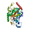
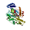
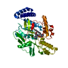
| Entry | Database: PDB / ID: 1wwj | |||||||||
|---|---|---|---|---|---|---|---|---|---|---|
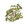
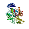
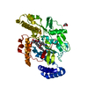
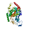
| Title | crystal structure of KaiB from Synechocystis sp. | |||||||||
 Components Components | Circadian clock protein kaiB | |||||||||
 Keywords Keywords | CIRCADIAN CLOCK PROTEIN / circadian / clock | |||||||||
| Function / homology |  Function and homology information Function and homology informationnegative regulation of phosphorylation / circadian rhythm / identical protein bindingSimilarity search - Function | |||||||||
| Biological species |  | |||||||||
| Method | X-RAY DIFFRACTION / SYNCHROTRON / MOLECULAR REPLACEMENT / Resolution: 1.9 Å | |||||||||
 Authors Authors | Hitomi, K. / Oyama, T. / Han, S. / Arvai, A.S. / Tainer, J.A. / Getzoff, E.D. | |||||||||
 Citation Citation | Journal: J.Biol.Chem. / Year: 2005 Title: Tetrameric architecture of the circadian clock protein KaiB. A novel interface for intermolecular interactions and its impact on the circadian rhythm. Authors: Hitomi, K. / Oyama, T. / Han, S. / Arvai, A.S. / Getzoff, E.D. | |||||||||
| History |
|
- Structure visualization
| Structure viewer | Molecule: MolmilJmol/JSmol |
|---|
- Downloads & links
-Download
| PDBx/mmCIF format | 1wwj.cif.gz | 97.5 KB | Display | PDBx/mmCIF format |
|---|---|---|---|---|
| PDB format | pdb1wwj.ent.gz | 73.9 KB | Display | PDB format |
| PDBx/mmJSON format | 1wwj.json.gz | Tree view | PDBx/mmJSON format | |
| Others |  Other downloads Other downloads |
-Validation report
| Arichive directory | https://data.pdbj.org/pub/pdb/validation_reports/ww/1wwjftp://data.pdbj.org/pub/pdb/validation_reports/ww/1wwj | HTTPS FTP |
|---|
-Related structure data
| Similar structure data |
|---|


-Links
 PDBj
PDBj


- Assembly
| Deposited unit | 
| ||||||||
|---|---|---|---|---|---|---|---|---|---|
| 1 |
| ||||||||
| Unit cell |
|
-Components
| #1: Protein | Mass: 11950.994 Da / Num. of mol.: 4 Source method: isolated from a genetically manipulated source Source: (gene. exp.) Escherichia coli (E. coli) / References: UniProt: P74645#2: Chemical | ChemComp-MLT / Malic acid  Mass: 134.087 Da / Num. of mol.: 8 / Source method: obtained synthetically / Formula: C4H6O5 Mass: 134.087 Da / Num. of mol.: 8 / Source method: obtained synthetically / Formula: C4H6O5#3: Chemical | ChemComp-BET / Trimethylglycine  Mass: 118.154 Da / Num. of mol.: 5 / Source method: obtained synthetically / Formula: C5H12NO2 Mass: 118.154 Da / Num. of mol.: 5 / Source method: obtained synthetically / Formula: C5H12NO2#4: Chemical | Imidazole  Mass: 69.085 Da / Num. of mol.: 3 / Source method: obtained synthetically / Formula: C3H5N2 Mass: 69.085 Da / Num. of mol.: 3 / Source method: obtained synthetically / Formula: C3H5N2#5: Water | ChemComp-HOH / | Water Mass: 18.015 Da / Num. of mol.: 237 / Source method: isolated from a natural source / Formula: H2O Mass: 18.015 Da / Num. of mol.: 237 / Source method: isolated from a natural source / Formula: H2O |
|---|
-Experimental details
-Experiment
| Experiment | Method: X-RAY DIFFRACTION / Number of used crystals: 1 |
|---|
- Sample preparation
| Crystal | Density Matthews: 2.1 Å3/Da / Density % sol: 41.3 % |
|---|---|
| Crystal grow | Temperature: 298 K / Method: vapor diffusion / pH: 6 Details: betaine, pH 6.0, VAPOR DIFFUSION, temperature 298.0K |
-Data collection
| Diffraction | Mean temperature: 100 K |
|---|---|
| Diffraction source | Source: SYNCHROTRON / Site: SSRL  / Beamline: BL7-1 / Wavelength: 1.08 Å / Beamline: BL7-1 / Wavelength: 1.08 Å |
| Detector | Type: MARRESEARCH / Detector: IMAGE PLATE / Date: May 15, 2000 |
| Radiation | Protocol: SINGLE WAVELENGTH / Monochromatic (M) / Laue (L): M / Scattering type: x-ray |
| Radiation wavelength | Wavelength: 1.08 Å / Relative weight: 1 |
| Reflection | Resolution: 1.9→30 Å / Num. obs: 31143 / % possible obs: 95.9 % / Observed criterion σ(F): 2 / Observed criterion σ(I): 2 / Biso Wilson estimate: 18.2 Å2 |
| Reflection shell | Resolution: 1.9→1.97 Å / % possible all: 89.9 |
- Processing
| Software |
| ||||||||||||||||||||||||||||||||||||||||||||||||||||||||||||||||||||||||||||||||
|---|---|---|---|---|---|---|---|---|---|---|---|---|---|---|---|---|---|---|---|---|---|---|---|---|---|---|---|---|---|---|---|---|---|---|---|---|---|---|---|---|---|---|---|---|---|---|---|---|---|---|---|---|---|---|---|---|---|---|---|---|---|---|---|---|---|---|---|---|---|---|---|---|---|---|---|---|---|---|---|---|---|
| Refinement | Method to determine structure: MOLECULAR REPLACEMENT / Resolution: 1.9→28.04 Å / Rfactor Rfree error: 0.007 / Data cutoff high absF: 1050233.55 / Data cutoff low absF: 0 / Isotropic thermal model: RESTRAINED / Cross valid method: THROUGHOUT / σ(F): 0
| ||||||||||||||||||||||||||||||||||||||||||||||||||||||||||||||||||||||||||||||||
| Solvent computation | Solvent model: FLAT MODEL / Bsol: 88.641 Å2 / ksol: 0.375667 e/Å3 | ||||||||||||||||||||||||||||||||||||||||||||||||||||||||||||||||||||||||||||||||
| Displacement parameters | Biso mean: 39.6 Å2
| ||||||||||||||||||||||||||||||||||||||||||||||||||||||||||||||||||||||||||||||||
| Refine analyze |
| ||||||||||||||||||||||||||||||||||||||||||||||||||||||||||||||||||||||||||||||||
| Refinement step | Cycle: LAST / Resolution: 1.9→28.04 Å
| ||||||||||||||||||||||||||||||||||||||||||||||||||||||||||||||||||||||||||||||||
| Refine LS restraints |
| ||||||||||||||||||||||||||||||||||||||||||||||||||||||||||||||||||||||||||||||||
| LS refinement shell | Resolution: 1.9→2.02 Å / Rfactor Rfree error: 0.023 / Total num. of bins used: 6
| ||||||||||||||||||||||||||||||||||||||||||||||||||||||||||||||||||||||||||||||||
| Xplor file |
|
