 Movie
Movie Controller

[English] 日本語
 Yorodumi
- PDB-1wvl: Crystal Structure of Multimeric DNA-binding Protein Sac7d-GCN4 wi... -
+ Open data

- Basic information
| Entry | Database: PDB / ID: 1wvl | ||||||
|---|---|---|---|---|---|---|---|
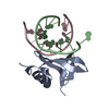
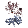
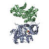
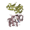
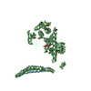
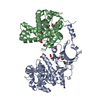
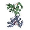
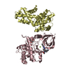
| Title | Crystal Structure of Multimeric DNA-binding Protein Sac7d-GCN4 with DNA decamer | ||||||
 Components Components |
| ||||||
 Keywords Keywords |  STRUCTURAL PROTEIN/DNA / leucine zipper / protein engineering / synchrotron radiation / thermophile / STRUCTURAL PROTEIN-DNA COMPLEX STRUCTURAL PROTEIN/DNA / leucine zipper / protein engineering / synchrotron radiation / thermophile / STRUCTURAL PROTEIN-DNA COMPLEX | ||||||
| Function / homology |  Function and homology information Function and homology information | ||||||
| Biological species |   Sulfolobus acidocaldarius (acidophilic) Sulfolobus acidocaldarius (acidophilic) | ||||||
| Method | X-RAY DIFFRACTION / MOLECULAR REPLACEMENT / Resolution: 2.6 Å | ||||||
 Authors Authors | Wu, S.W. / Wang, A.H. | ||||||
 Citation Citation | Journal: Proteins / Year: 2005 Title: Design and characterization of a multimeric DNA binding protein using Sac7d and GCN4 as templates Authors: Wu, S.W. / Ko, T.P. / Chou, C.C. / Wang, A.H. | ||||||
| History |
|
- Structure visualization
| Structure viewer | Molecule: MolmilJmol/JSmol |
|---|
- Downloads & links
-Download
| PDBx/mmCIF format | 1wvl.cif.gz | 60.5 KB | Display | PDBx/mmCIF format |
|---|---|---|---|---|
| PDB format | pdb1wvl.ent.gz | 41.9 KB | Display | PDB format |
| PDBx/mmJSON format | 1wvl.json.gz | Tree view | PDBx/mmJSON format | |
| Others |  Other downloads Other downloads |
-Validation report
| Arichive directory | https://data.pdbj.org/pub/pdb/validation_reports/wv/1wvlftp://data.pdbj.org/pub/pdb/validation_reports/wv/1wvl | HTTPS FTP |
|---|
-Related structure data
| Related structure data |  1azpS S: Starting model for refinement |
|---|---|
| Similar structure data |


-Links
 PDBj
PDBj- Assembly
| Deposited unit | 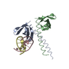
| ||||||||
|---|---|---|---|---|---|---|---|---|---|
| 1 | 
| ||||||||
| Unit cell |
| ||||||||
| Components on special symmetry positions |
| ||||||||
| Details | The biological assembly is a tetramer generated from the dimer in the asymmetric unit by the operations: -1+y, 1+x, 3/4-z. |
-Components
| #1: DNA chain | Mass: 3044.016 Da / Num. of mol.: 2 / Source method: obtained synthetically #2: Protein | Mass: 9218.810 Da / Num. of mol.: 2 Source method: isolated from a genetically manipulated source Source: (gene. exp.) Sulfolobus acidocaldarius (acidophilic)Plasmid: pET-11a / Species (production host): Escherichia coli / Production host:  Escherichia coli BL21(DE3) (bacteria) / Strain (production host): BL21 (DE3) / References: UniProt: P13123 Escherichia coli BL21(DE3) (bacteria) / Strain (production host): BL21 (DE3) / References: UniProt: P13123#3: Water | ChemComp-HOH / | Water Mass: 18.015 Da / Num. of mol.: 146 / Source method: isolated from a natural source / Formula: H2O Mass: 18.015 Da / Num. of mol.: 146 / Source method: isolated from a natural source / Formula: H2O |
|---|
-Experimental details
-Experiment
| Experiment | Method: X-RAY DIFFRACTION / Number of used crystals: 2 |
|---|
- Sample preparation
| Crystal | Density Matthews: 3.27 Å3/Da / Density % sol: 62.35 % | ||||||||||||||||||||
|---|---|---|---|---|---|---|---|---|---|---|---|---|---|---|---|---|---|---|---|---|---|
| Crystal grow | Temperature: 298 K / Method: vapor diffusion, hanging drop / pH: 6.5 Details: 1.6M Tri-Sodium Citrate, pH 6.5, VAPOR DIFFUSION, HANGING DROP, temperature 298K | ||||||||||||||||||||
| Components of the solutions |
|
-Data collection
| Diffraction | Mean temperature: 116 K |
|---|---|
| Diffraction source | Source: ROTATING ANODE / Type: RIGAKU MICROMAX-007 / Wavelength: 1.5418 Å |
| Detector | Type: RIGAKU RAXIS IV++ / Detector: IMAGE PLATE / Date: Apr 30, 2003 |
| Radiation | Monochromator: MSC confocal optics / Protocol: SINGLE WAVELENGTH / Monochromatic (M) / Laue (L): M / Scattering type: x-ray |
| Radiation wavelength | Wavelength: 1.5418 Å / Relative weight: 1 |
| Reflection | Resolution: 2.6→20 Å / Num. all: 11603 / Num. obs: 10690 / % possible obs: 99.5 % / Observed criterion σ(F): 2 / Observed criterion σ(I): 1 |
| Reflection shell | Resolution: 2.6→2.7 Å / % possible all: 100 |
- Processing
| Software |
| ||||||||||||||||||||
|---|---|---|---|---|---|---|---|---|---|---|---|---|---|---|---|---|---|---|---|---|---|
| Refinement | Method to determine structure: MOLECULAR REPLACEMENT Starting model: 1azp Resolution: 2.6→20 Å / Cross valid method: THROUGHOUT / σ(F): 3 / Stereochemistry target values: Engh & Huber
| ||||||||||||||||||||
| Refine analyze |
| ||||||||||||||||||||
| Refinement step | Cycle: LAST / Resolution: 2.6→20 Å
| ||||||||||||||||||||
| Refine LS restraints |
| ||||||||||||||||||||
| LS refinement shell | Resolution: 2.6→2.76 Å / Rfactor Rfree error: 0.046
|
