 Movie
Movie Controller
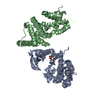


+ Open data

- Basic information
| Entry | Database: PDB / ID: 1wsr | ||||||
|---|---|---|---|---|---|---|---|
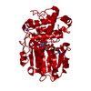
| Title | Crystal Structure of Human T-protein of Glycine Cleavage System | ||||||
 Components Components | Aminomethyltransferase | ||||||
 Keywords Keywords | TRANSFERASE / GLYCINE-CLEAVAGE SYTEM / AMINOMETHYL TRANSFERASE | ||||||
| Function / homology |  Function and homology informationaminomethyltransferase / glycine catabolic process / aminomethyltransferase activity / Glycine degradation / glycine decarboxylation via glycine cleavage system / transaminase activity / mitochondrial matrix / mitochondrion / nucleoplasm Function and homology informationaminomethyltransferase / glycine catabolic process / aminomethyltransferase activity / Glycine degradation / glycine decarboxylation via glycine cleavage system / transaminase activity / mitochondrial matrix / mitochondrion / nucleoplasmSimilarity search - Function | ||||||
| Biological species |  Homo sapiens (human) Homo sapiens (human) | ||||||
| Method | X-RAY DIFFRACTION / SYNCHROTRON / SAD / Resolution: 2 Å | ||||||
 Authors Authors | Okamura-Ikeda, K. / Hosaka, H. / Yoshimura, M. / Yamashita, E. / Toma, S. / Nakagawa, A. / Fujiwara, K. / Motokawa, Y. / Taniguchi, H. | ||||||
 Citation Citation | Journal: J.Mol.Biol. / Year: 2005 Title: Crystal Structure of Human T-protein of Glycine Cleavage System at 2.0A Resolution and its Implication for Understanding Non-ketotic Hyperglycinemia Authors: Okamura-Ikeda, K. / Hosaka, H. / Yoshimura, M. / Yamashita, E. / Toma, S. / Nakagawa, A. / Fujiwara, K. / Motokawa, Y. / Taniguchi, H. | ||||||
| History |
|
- Structure visualization
| Structure viewer | Molecule: MolmilJmol/JSmol |
|---|
- Downloads & links
-Download
| PDBx/mmCIF format | 1wsr.cif.gz | 178.5 KB | Display | PDBx/mmCIF format |
|---|---|---|---|---|
| PDB format | pdb1wsr.ent.gz | 139.1 KB | Display | PDB format |
| PDBx/mmJSON format | 1wsr.json.gz | Tree view | PDBx/mmJSON format | |
| Others |  Other downloads Other downloads |
-Validation report
| Arichive directory | https://data.pdbj.org/pub/pdb/validation_reports/ws/1wsrftp://data.pdbj.org/pub/pdb/validation_reports/ws/1wsr | HTTPS FTP |
|---|
-Related structure data


-Links
 PDBj
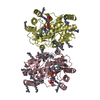
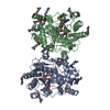
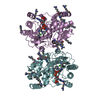
PDBj- Assembly
| Deposited unit | 
| ||||||||
|---|---|---|---|---|---|---|---|---|---|
| 1 |
| ||||||||
| 2 | 
| ||||||||
| Unit cell |
|
-Components
| #1: Protein | / Glycine cleavage system T protein / GCVT Mass: 40928.219 Da / Num. of mol.: 2 Source method: isolated from a genetically manipulated source Source: (gene. exp.) Homo sapiens (human) / Gene: GCST / Plasmid: pET3 / Production host:  Escherichia coli (E. coli) / Strain (production host): BL21(DE3)pLysS / References: UniProt: P48728, aminomethyltransferase Escherichia coli (E. coli) / Strain (production host): BL21(DE3)pLysS / References: UniProt: P48728, aminomethyltransferase#2: Chemical | ChemComp-SO4 / Sulfate  Mass: 96.063 Da / Num. of mol.: 11 / Source method: obtained synthetically / Formula: SO4 Mass: 96.063 Da / Num. of mol.: 11 / Source method: obtained synthetically / Formula: SO4#3: Water | ChemComp-HOH / | Water Mass: 18.015 Da / Num. of mol.: 1177 / Source method: isolated from a natural source / Formula: H2O Mass: 18.015 Da / Num. of mol.: 1177 / Source method: isolated from a natural source / Formula: H2O |
|---|
-Experimental details
-Experiment
| Experiment | Method: X-RAY DIFFRACTION / Number of used crystals: 1 |
|---|
- Sample preparation
| Crystal | Density Matthews: 2.9 Å3/Da / Density % sol: 57.2 % |
|---|---|
| Crystal grow | Temperature: 298 K / Method: vapor diffusion, hanging drop / pH: 5.6 Details: ammonium sulfate, potassium sodium tartrate, citrate, pH 5.6, VAPOR DIFFUSION, HANGING DROP, temperature 298K |
-Data collection
| Diffraction | Mean temperature: 100 K |
|---|---|
| Diffraction source | Source: SYNCHROTRON / Site: SPring-8  / Beamline: BL44XU / Wavelength: 0.9 Å / Beamline: BL44XU / Wavelength: 0.9 Å |
| Detector | Type: Bruker DIP-6040 / Detector: CCD / Date: May 31, 2003 |
| Radiation | Monochromator: Si(111) / Protocol: SINGLE WAVELENGTH / Monochromatic (M) / Laue (L): M / Scattering type: x-ray |
| Radiation wavelength | Wavelength: 0.9 Å / Relative weight: 1 |
| Reflection | Resolution: 2→95.3 Å / Num. all: 64914 / Num. obs: 64914 / % possible obs: 100 % / Observed criterion σ(F): 0 / Observed criterion σ(I): 0 / Redundancy: 8.5 % / Biso Wilson estimate: 9 Å2 / Rmerge(I) obs: 0.096 |
| Reflection shell | Resolution: 2→2.07 Å / Redundancy: 8.2 % / Rmerge(I) obs: 0.352 / Mean I/σ(I) obs: 9.9 / Num. unique all: 6380 / % possible all: 100 |
- Processing
| Software |
| ||||||||||||||||||||||||||||||||||||
|---|---|---|---|---|---|---|---|---|---|---|---|---|---|---|---|---|---|---|---|---|---|---|---|---|---|---|---|---|---|---|---|---|---|---|---|---|---|
| Refinement | Method to determine structure: SAD / Resolution: 2→49.92 Å / Rfactor Rfree error: 0.003 / Data cutoff high absF: 2538972.03 / Data cutoff low absF: 0 / Isotropic thermal model: RESTRAINED / Cross valid method: THROUGHOUT / σ(F): 0 / Stereochemistry target values: Engh & Huber
| ||||||||||||||||||||||||||||||||||||
| Solvent computation | Solvent model: FLAT MODEL / Bsol: 62.3929 Å2 / ksol: 0.378175 e/Å3 | ||||||||||||||||||||||||||||||||||||
| Displacement parameters | Biso mean: 20.6 Å2
| ||||||||||||||||||||||||||||||||||||
| Refine analyze |
| ||||||||||||||||||||||||||||||||||||
| Refinement step | Cycle: LAST / Resolution: 2→49.92 Å
| ||||||||||||||||||||||||||||||||||||
| Refine LS restraints |
| ||||||||||||||||||||||||||||||||||||
| LS refinement shell | Resolution: 2→2.13 Å / Rfactor Rfree error: 0.009 / Total num. of bins used: 6
| ||||||||||||||||||||||||||||||||||||
| Xplor file |
|
