 Movie
Movie Controller

[English] 日本語
 Yorodumi
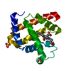
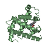
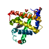
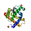
- PDB-1wsh: Crystal structure of E.coli RNase HI active site mutant (E48A/K87A) -
+ Open data

- Basic information
| Entry | Database: PDB / ID: 1wsh | ||||||
|---|---|---|---|---|---|---|---|
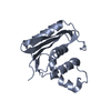
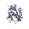
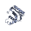
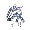
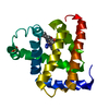
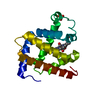
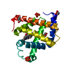
| Title | Crystal structure of E.coli RNase HI active site mutant (E48A/K87A) | ||||||
 Components Components | Ribonuclease HI | ||||||
 Keywords Keywords |  HYDROLASE / RNase H / active-site mutant HYDROLASE / RNase H / active-site mutant | ||||||
| Function / homology |  Function and homology informationDNA replication, removal of RNA primer / ribonuclease H / RNA-DNA hybrid ribonuclease activity / endonuclease activity / nucleic acid binding / magnesium ion binding / cytoplasm Function and homology informationDNA replication, removal of RNA primer / ribonuclease H / RNA-DNA hybrid ribonuclease activity / endonuclease activity / nucleic acid binding / magnesium ion binding / cytoplasmSimilarity search - Function | ||||||
| Biological species |  Escherichia coli (E. coli) Escherichia coli (E. coli) | ||||||
| Method | X-RAY DIFFRACTION / SYNCHROTRON / MOLECULAR REPLACEMENT / Resolution: 1.9 Å | ||||||
 Authors Authors | Tsunaka, Y. / Takano, K. / Matsumura, H. / Yamagata, Y. / Kanaya, S. | ||||||
| History |
|
- Structure visualization
| Structure viewer | Molecule: MolmilJmol/JSmol |
|---|
- Downloads & links
-Download
| PDBx/mmCIF format | 1wsh.cif.gz | 131 KB | Display | PDBx/mmCIF format |
|---|---|---|---|---|
| PDB format | pdb1wsh.ent.gz | 104.6 KB | Display | PDB format |
| PDBx/mmJSON format | 1wsh.json.gz | Tree view | PDBx/mmJSON format | |
| Others |  Other downloads Other downloads |
-Validation report
| Arichive directory | https://data.pdbj.org/pub/pdb/validation_reports/ws/1wshftp://data.pdbj.org/pub/pdb/validation_reports/ws/1wsh | HTTPS FTP |
|---|
-Related structure data
| Related structure data | |
|---|---|
| Similar structure data |


-Links
 PDBj
PDBj
- Assembly
| Deposited unit | 
| ||||||||
|---|---|---|---|---|---|---|---|---|---|
| 1 | 
| ||||||||
| 2 | 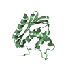
| ||||||||
| 3 | 
| ||||||||
| 4 | 
| ||||||||
| Unit cell |
|
-Components
| #1: Protein | Mass: 17506.859 Da / Num. of mol.: 4 / Mutation: E48A/K87A Source method: isolated from a genetically manipulated source Source: (gene. exp.) Escherichia coli (E. coli) / Plasmid: pJAL600 / Production host: Escherichia coli (E. coli) / References: UniProt: P0A7Y4, ribonuclease H#2: Water | ChemComp-HOH / | Water Mass: 18.015 Da / Num. of mol.: 346 / Source method: isolated from a natural source / Formula: H2O Mass: 18.015 Da / Num. of mol.: 346 / Source method: isolated from a natural source / Formula: H2O |
|---|
-Experimental details
-Experiment
| Experiment | Method: X-RAY DIFFRACTION / Number of used crystals: 1 |
|---|
- Sample preparation
| Crystal | Density Matthews: 2.24 Å3/Da / Density % sol: 45.05 % |
|---|
-Data collection
| Diffraction source | Source: SYNCHROTRON / Site: SPring-8  / Beamline: BL44XU / Wavelength: 0.9 Å / Beamline: BL44XU / Wavelength: 0.9 Å |
|---|---|
| Detector | Type: MACSCIENCE / Detector: IMAGE PLATE / Date: Jun 21, 2003 |
| Radiation | Protocol: SINGLE WAVELENGTH / Monochromatic (M) / Laue (L): M / Scattering type: x-ray |
| Radiation wavelength | Wavelength: 0.9 Å / Relative weight: 1 |
| Reflection | Resolution: 1.9→50 Å / Num. all: 50326 / Num. obs: 50229 / % possible obs: 99.8 % |
| Reflection shell | Highest resolution: 1.9 Å |
- Processing
| Software | Name: CNS / Classification: refinement | |||||||||||||||
|---|---|---|---|---|---|---|---|---|---|---|---|---|---|---|---|---|
| Refinement | Method to determine structure: MOLECULAR REPLACEMENT / Resolution: 1.9→50 Å
| |||||||||||||||
| Refinement step | Cycle: LAST / Resolution: 1.9→50 Å
|
