 Movie
Movie Controller


+ Open data

- Basic information
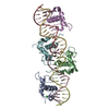
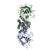
| Entry | Database: PDB / ID: 1wdc | ||||||
|---|---|---|---|---|---|---|---|
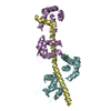
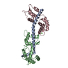
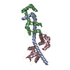
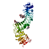
| Title | SCALLOP MYOSIN REGULATORY DOMAIN | ||||||
 Components Components | (SCALLOP MYOSIN) x 3 | ||||||
 Keywords Keywords |  MUSCLE PROTEIN / MYOSIN / CALCIUM BINDING PROTEIN MUSCLE PROTEIN / MYOSIN / CALCIUM BINDING PROTEIN | ||||||
| Function / homology |  Function and homology information Function and homology informationmyosin filament / myosin complex / myofibril / cytoskeletal motor activity / actin filament binding / calmodulin binding / calcium ion binding / ATP bindingSimilarity search - Function | ||||||
| Biological species |  Argopecten irradians (bay scallop) Argopecten irradians (bay scallop) | ||||||
| Method | X-RAY DIFFRACTION / Resolution: 2 Å | ||||||
 Authors Authors | Houdusse, A. / Cohen, C. | ||||||
 Citation Citation | Journal: Structure / Year: 1996 Title: Structure of the regulatory domain of scallop myosin at 2 A resolution: implications for regulation. Authors: Houdusse, A. / Cohen, C. #1: Journal: Nature / Year: 1994Title: Structure of the Regulatory Domain of Scallop Myosin at 2.8 A Resolution Authors: Xie, X. / Harrison, D.H. / Schlichting, I. / Sweet, R.M. / Kalabokis, V.N. / Szent-Gyorgyi, A.G. / Cohen, C. #2: Journal: Proc.Natl.Acad.Sci.USA / Year: 1990Title: Isolation of the Regulatory Domain of Scallop Myosin: Role of the Essential Light Chain in Calcium Binding Authors: Kwon, H. / Goodwin, E.B. / Nyitray, L. / Berliner, E. / O'Neall-Hennessey, E. / Melandri, F.D. / Szent-Gyorgyi, A.G. | ||||||
| History |
|
- Structure visualization
| Structure viewer | Molecule: MolmilJmol/JSmol |
|---|
- Downloads & links
-Download
| PDBx/mmCIF format | 1wdc.cif.gz | 90 KB | Display | PDBx/mmCIF format |
|---|---|---|---|---|
| PDB format | pdb1wdc.ent.gz | 67 KB | Display | PDB format |
| PDBx/mmJSON format | 1wdc.json.gz | Tree view | PDBx/mmJSON format | |
| Others |  Other downloads Other downloads |
-Validation report
| Arichive directory | https://data.pdbj.org/pub/pdb/validation_reports/wd/1wdcftp://data.pdbj.org/pub/pdb/validation_reports/wd/1wdc | HTTPS FTP |
|---|
-Related structure data
| Similar structure data |
|---|


-Links
 PDBj
PDBj


- Assembly
| Deposited unit | 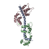
| ||||||||
|---|---|---|---|---|---|---|---|---|---|
| 1 |
| ||||||||
| Unit cell |
|
-Components
-Protein , 3 types, 3 molecules ABC
| #1: Protein | Mass: 7997.623 Da / Num. of mol.: 1 / Fragment: PROTEOLYTIC FRAGMENT, REGULATORY DOMAIN / Source method: isolated from a natural source / Details: PH 7.0 / Source: (natural) Argopecten irradians (bay scallop) / Organ: SKELETALSkeleton / Tissue: SKELETAL MUSCLE / References: UniProt: P24733 |
|---|---|
| #2: Protein | Mass: 17560.855 Da / Num. of mol.: 1 / Fragment: PROTEOLYTIC FRAGMENT, REGULATORY DOMAIN / Source method: isolated from a natural source / Details: PH 7.0 / Source: (natural) Argopecten irradians (bay scallop) / Organ: SKELETALSkeleton / Tissue: SKELETAL MUSCLE / References: UniProt: P13543 |
| #3: Protein | Mass: 17635.635 Da / Num. of mol.: 1 / Fragment: PROTEOLYTIC FRAGMENT, REGULATORY DOMAIN / Source method: isolated from a natural source / Details: PH 7.0 / Source: (natural) Argopecten irradians (bay scallop) / Organ: SKELETALSkeleton / Tissue: SKELETAL MUSCLE / References: UniProt: P07291 |
-Non-polymers , 3 types, 166 molecules 


| #4: Chemical | ChemComp-MG /  Mass: 24.305 Da / Num. of mol.: 1 / Source method: obtained synthetically / Formula: Mg Mass: 24.305 Da / Num. of mol.: 1 / Source method: obtained synthetically / Formula: Mg |
|---|---|
| #5: Chemical | ChemComp-CA /  Mass: 40.078 Da / Num. of mol.: 1 / Source method: obtained synthetically / Formula: Ca Mass: 40.078 Da / Num. of mol.: 1 / Source method: obtained synthetically / Formula: Ca |
| #6: Water | ChemComp-HOH / WaterMass: 18.015 Da / Num. of mol.: 164 / Source method: isolated from a natural source / Formula: H2O |
-Details
| Compound details | THE RD FRAGMENT IS A TERNARY COMPLEX OF A PORTION OF HEAVY CHAIN WITH TWO LIGHT CHAINS. CHAIN LABEL ...THE RD FRAGMENT IS A TERNARY COMPLEX OF A PORTION OF HEAVY CHAIN WITH TWO LIGHT CHAINS. CHAIN LABEL A DESIGNATES |
|---|
-Experimental details
-Experiment
| Experiment | Method: X-RAY DIFFRACTION |
|---|
- Sample preparation
| Crystal | Density Matthews: 2.68 Å3/Da / Density % sol: 51.9 % | ||||||||||||||||||||||||||||||||||||||||||
|---|---|---|---|---|---|---|---|---|---|---|---|---|---|---|---|---|---|---|---|---|---|---|---|---|---|---|---|---|---|---|---|---|---|---|---|---|---|---|---|---|---|---|---|
| Crystal grow | pH: 7 / Details: pH 7.0 | ||||||||||||||||||||||||||||||||||||||||||
| Crystal grow | *PLUS Temperature: 4 ℃ / Method: vapor diffusion, hanging drop / Details: Xie, X., (1994) Nature, 368, 306. | ||||||||||||||||||||||||||||||||||||||||||
| Components of the solutions | *PLUS
|
-Data collection
| Diffraction | Mean temperature: 277 K |
|---|---|
| Diffraction source | Wavelength: 1.5418 |
| Detector | Type: RIGAKU / Detector: IMAGE PLATE / Date: Oct 1, 1993 |
| Radiation | Monochromatic (M) / Laue (L): M / Scattering type: x-ray |
| Radiation wavelength | Wavelength: 1.5418 Å / Relative weight: 1 |
| Reflection | Resolution: 2.02→100 Å / Num. obs: 26715 / % possible obs: 89.2 % / Rmerge(I) obs: 0.061 |
| Reflection | *PLUS Num. obs: 26725 / Num. measured all: 104023 |
- Processing
| Software |
| ||||||||||||||||||||||||||||||||||||||||||||||||||||||||||||
|---|---|---|---|---|---|---|---|---|---|---|---|---|---|---|---|---|---|---|---|---|---|---|---|---|---|---|---|---|---|---|---|---|---|---|---|---|---|---|---|---|---|---|---|---|---|---|---|---|---|---|---|---|---|---|---|---|---|---|---|---|---|
| Refinement | Resolution: 2→8 Å / σ(F): 2 Details: SIDE CHAIN ATOMS OF RESIDUE GLU B 153 ARE NOT SEEN IN THE ELECTRON DENSITY.
| ||||||||||||||||||||||||||||||||||||||||||||||||||||||||||||
| Displacement parameters | Biso mean: 36.52 Å2 | ||||||||||||||||||||||||||||||||||||||||||||||||||||||||||||
| Refine analyze | Luzzati coordinate error obs: 0.2 Å | ||||||||||||||||||||||||||||||||||||||||||||||||||||||||||||
| Refinement step | Cycle: LAST / Resolution: 2→8 Å
| ||||||||||||||||||||||||||||||||||||||||||||||||||||||||||||
| Refine LS restraints |
| ||||||||||||||||||||||||||||||||||||||||||||||||||||||||||||
| Software | *PLUS Name: X-PLOR / Version: 3.1 / Classification: refinement | ||||||||||||||||||||||||||||||||||||||||||||||||||||||||||||
| Refine LS restraints | *PLUS Type: x_dihedral_angle_d / Dev ideal: 24.135 |
